The effect of learning on neural activity in visual cortex and higher level brain areas
Animals need to continuously learn which sensory objects in their environment are behaviourally relevant because they predict rewards or dangers and can guide our actions. My lab is interested in how activity in early visual cortex and higher level areas changes when animals learn the significance of sensory stimuli. We use a virtual reality environment in which animals actively interact with visual stimuli and quickly learn which visual stimuli predict rewards and which do not. We use 2-photon calcium imaging to measure activity in large groups of cells while animals learn, so that we can compare responses of the same cells before and after learning. We find that representations of task-relevant stimuli become increasingly distinguishable with learning due to stabilization of existing and recruitment of new neurons. In addition, we find that signals emerge with learning that reflect anticipation and the behavioural choice of the animals.
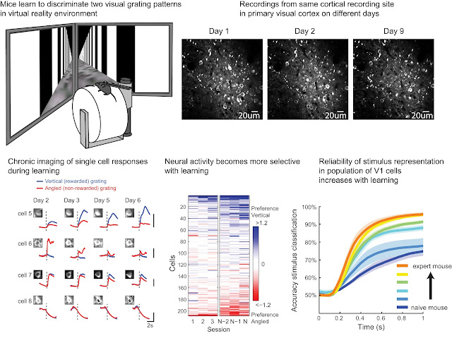 |
Figure 1. See also 'Learning Enhances Sensory and Multiple Nonsensory Representation in Primary Visual Cortex', Poort, Khan, Pachitariu, Nemri, Orsolic, Krupic, Bauza, Sahani, Keller, Mrsic-Flogel, Hofer, Neuron 2015. |
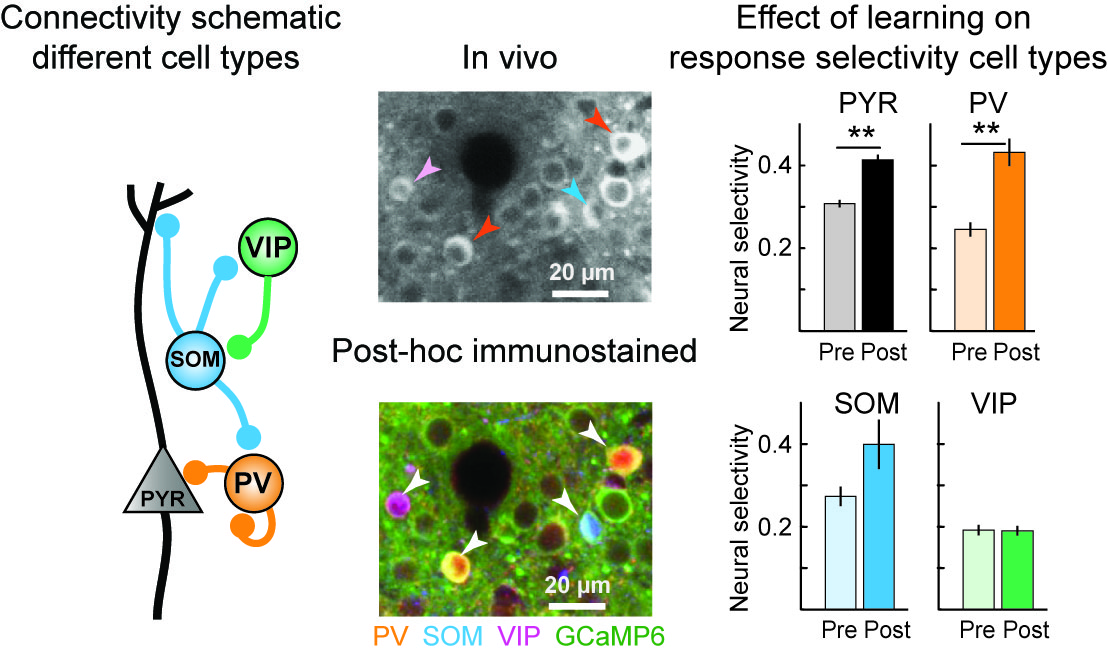
To probe the detailed circuits for learning, we have started to investigate the role of different cell types in the circuit. We use techniques that allow us to identify the cell type of cells imaged in-vivo, by post-hoc immunostaining and image registration to link stained brain sections to previously recorded in-vivo images (Fig 2, middle). This approach reveals for example that learning has particularly strong effects on PV interneurons (Fig 2, right).
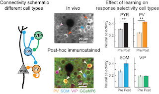 |
| Figure 2. See also 'Distinct learning-induced changes in stimulus selectivity and interactions of GABAergic interneuron classes in visual cortex’, Khan, Poort, Chadwick, Blot, Sahani, Mrsic-Flogel, Hofer, Nature Neuroscience 2018. |
Neural correlates of perceptual organization and task-dependent attention
We want to understand how our brain organizes the continuous bombardment by incoming sensory input into coherent percepts. One fundamental processing step is the assignment of image elements to the foreground ('figure') or background. With the use of electrophysiological recordings in primary visual cortex and higher level areas, we study how cells in these areas respond to different image elements.
Even in primary visual cortex, neurons respond more strongly to image elements that belong to a figure compared to background elements (Lamme et al., J Neurosci, 1995). However, image segmentation in the brain can depend on whether we attend to objects or not. We found that the detection of figure boundaries occurs early and depends little on attention, whereas filling in of the centre of the figure occurs later and is strongly enhanced by attention (see Figure 3, Poort et al., Neuron, 2012, see also Poort et al., Cereb Cortex 2016).
In the visual discrimination task in the virtual reality environment (Figure 1), responses become more selective with learning, but in addition, they also depend strongly on whether the mouse is attending to visual objects or not (Poort, Khan et al., Neuron 2015). One important aim of the lab is to establish the circuit-mechanisms of this flexible 'short-term' task-dependent attentional modulation (also in comparison to long-term learning-related response enhancements).
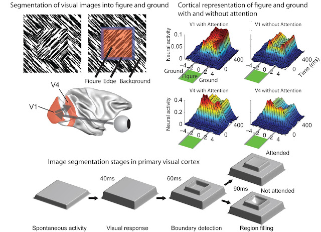 |
Figure 3. See also Poort, Raudies, Wannig, Lamme, Neumann, Roelfsema, Neuron 2012. |
Population coding of sensory stimulus features and modulation by learning and task-switching
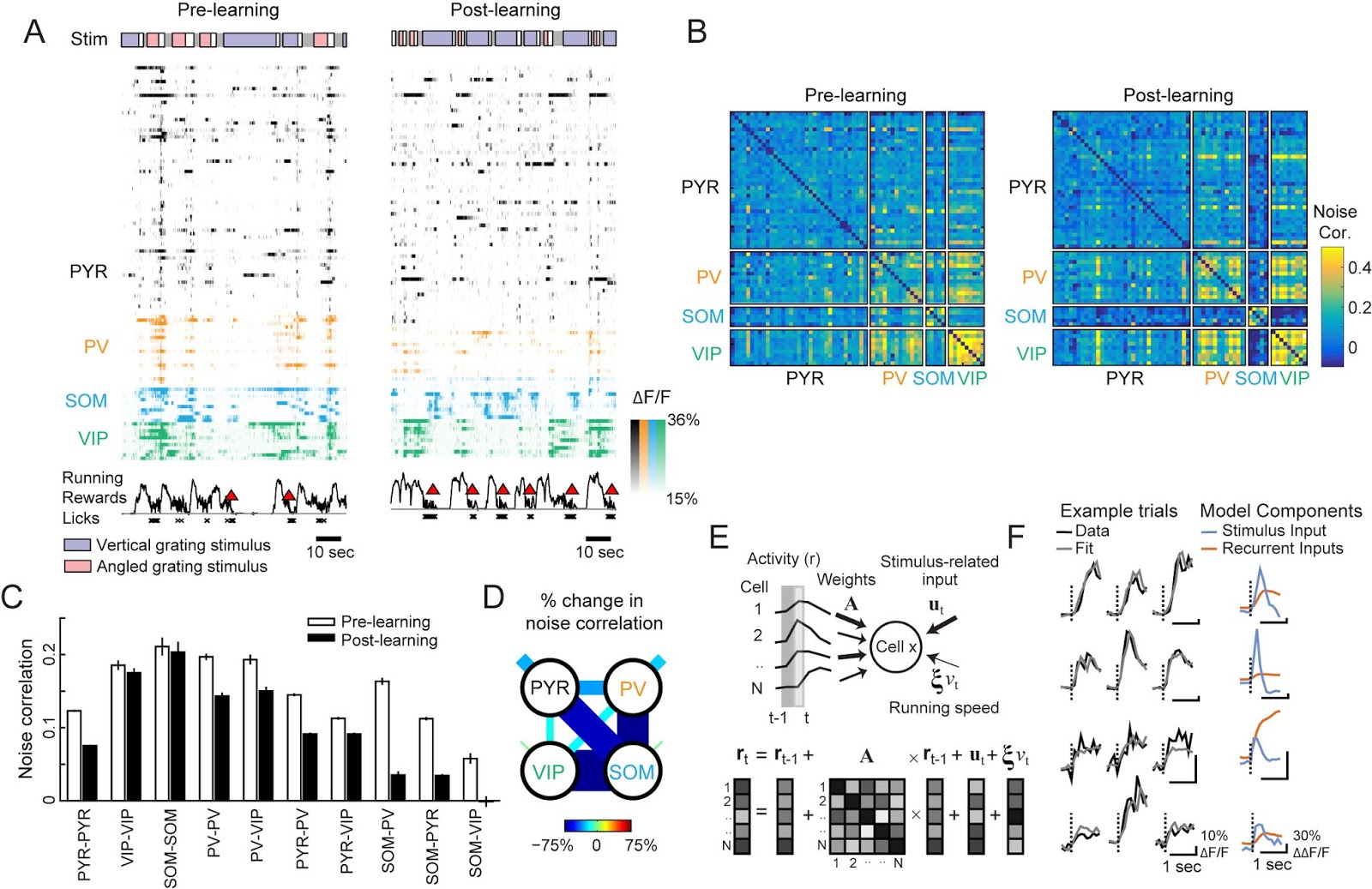
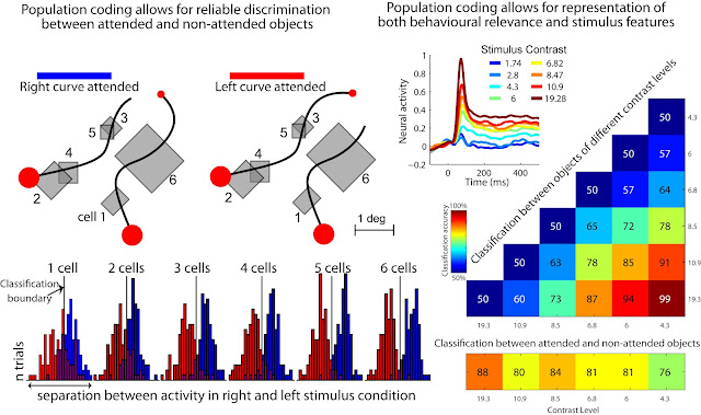
Complex cognitive functions rely on coordinated activity of groups of cells within different areas. We are interested in how populations of cells represent multiple types of information about sensory stimuli (see Figure 1 bottom right panel, and Figure 3) and how learning and changing task-requirements modify the interactions between populations of cells (Fig 4). We collaborate with computational neuroscientists to develop advanced models of complex neural circuits that can capture the contributions of different cell types and other influences on response properties (see Fig 4).
|
| Figure 4. See also Poort and Roelfsema, Cereb Ctx, 2009, and Pooresmaeili, Poort, Thiele, Roelfsema, J Neurosci, 2011. |
 |
| Figure 5. See also Khan Poort et al., Nature Neuroscience 2018. A. Example simultaneous recordings from multiple cell types B-D. Cell-type specific changes in response correlations during learning. E,F Multivariate autoregressive (MVAR) linear dynamical system model (A. Chadwick, M. Sahani, Gatsby Computational Neuroscience Unit) to fit responses of individual cells based on their interactions with simultaneously imaged cells. |
Translation of research results: from controlled experimental settings to more unrestrained natural conditions, and from mouse to man.
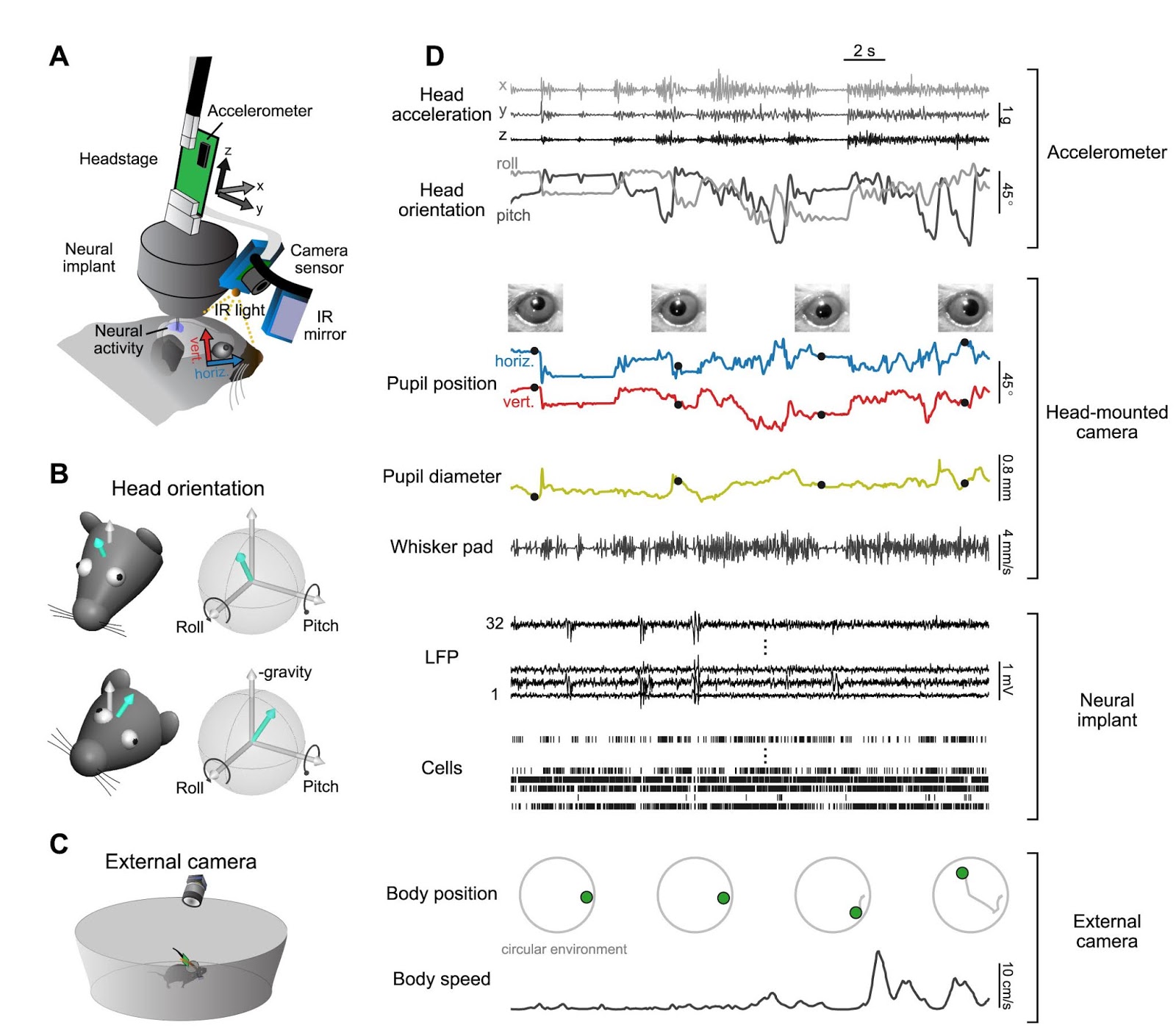
An important long-term goal of the lab is to relate advances in our understanding of circuits for sensory selection in mice to disease models and clinical populations. We are therefore collaborating with multiple labs in Cambridge who study mouse models of neurodevelopmental disorders associated with sensory selection deficits, such as schizophrenia and autism. For example, we study a mouse model of 22q11.2 deletion syndrome (the biggest known genetic risk factor for schizophrenia) to compare circuits for successful and unsuccessful sensory selection. Much behavioural work on cognitive function in mouse models is currently performed in freely moving animals. However, one challenge in visual neuroscience is that it has not been possible so far to monitor eye position in freely moving mice. We have therefore recently developed new methods to overcome this problem and combine detailed behavioural tracking with electrophysiology in freely moving mice (Fig. 6).
In order to relate findings in mice and men, we collaborate with groups in Cambridge that work on visual learning and attention in human (clinical) populations.
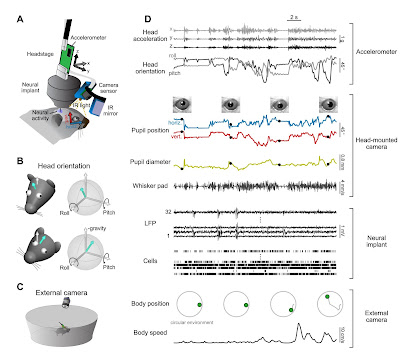 |
Figure 6. See Meyer, Poort, O'Keefe, Sahani, and Linden, Neuron 2018. A head-mounted camera system integrates detailed behavioral monitoring (e.g. eye, head, whisker movment) with multichannel electrophysiology in freely moving mice. |