Department of Physiology, Development and Neuroscience
Cranium, general and pathological collection
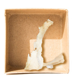
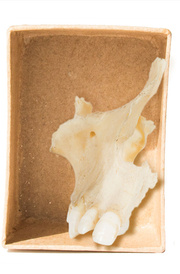
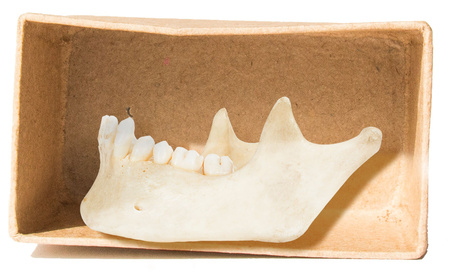
Right maxilla
Adult right maxilla, anterior view. The maxillae are a pair of bones that form the upper jaw, a large part of the lower face, and the nasal aperture. The two bones join at the midline of the hard palate. On the 360-degree image the maxillary sinus can be seen. This is a large void, which serves to reduce the skull’s weight, produce mucus and contribute to the quality of the voice.
Orientation: anterior view
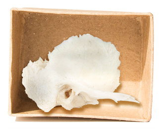
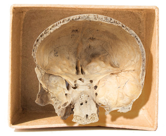
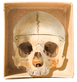
Cranium with metopic suture
Anterior view of adult cranium, demonstrating persistence of metopic suture of the frontal bone. The metopic suture is usually not visible after childhood, but occasionally persists into adulthood as in this case.
In life, sutures are held together by collagen fibres. At birth, the frontal bone is composed of two symmetrical halves, separated by the metopic suture. Generally, this fuses completely by the end of the second year of life. If persistent, then the frontal bone may show a greater degree of curvature.
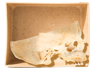
Median section through cranium, left half
Adult cranium (mandible and part of the calvaria removed), divided in the sagittal plane. Note some post-mortem damage to the bony orbit and lateral wall of the nasal cavity, and to the dentition.
The cranium is the most complicated skeletal structure. It contains the brain and organs of sight, smell and hearing, and the entry points to the respiratory and alimentary systems. Visible in this image are the sinuses, or air-filled cavities of the frontal bone and maxilla, and the crista galli (‘coxcomb’), an elevated ridge that projects from the cribriform plate of the ethmoid bone. The spongy bone separating the inner and outer layers of the cortical bone of the skull seen in this image is called ‘diploe’.
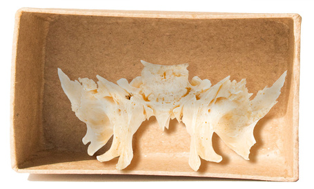
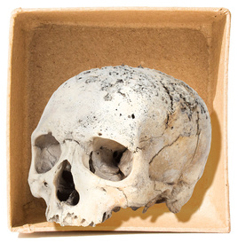
Cranium showing osseous destruction: syphilis
Adult cranium in two parts (coronal section) demonstrating areas of erosion and remodelling, particularly prominent across the superior aspect of the frontal bone and parietal bones.
The combination of osteolytic lesions, remodelling and scarring in the skull is known is caries sicca, and is pathognomonic of treponematosis (acquired/endemic syphilis, yaws).
Pathology: treponematosis, caries sicca
Orientation: anterior
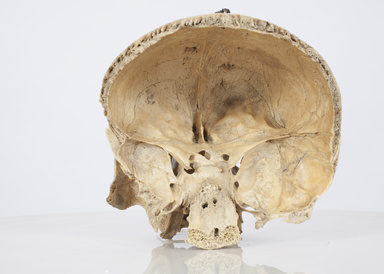
Partial cranium, coronal section
Coronal section of adult skull, demonstrating the viscerocranium (facial skeleton). Parts of the delicate ethmoid and lacrimal bones in the medial orbital walls have been damaged.
On the 360-degree image, the anterior and middle cranial fossae can be seen, together with impressions of meningeal blood vessels on the inner surface of the skull.