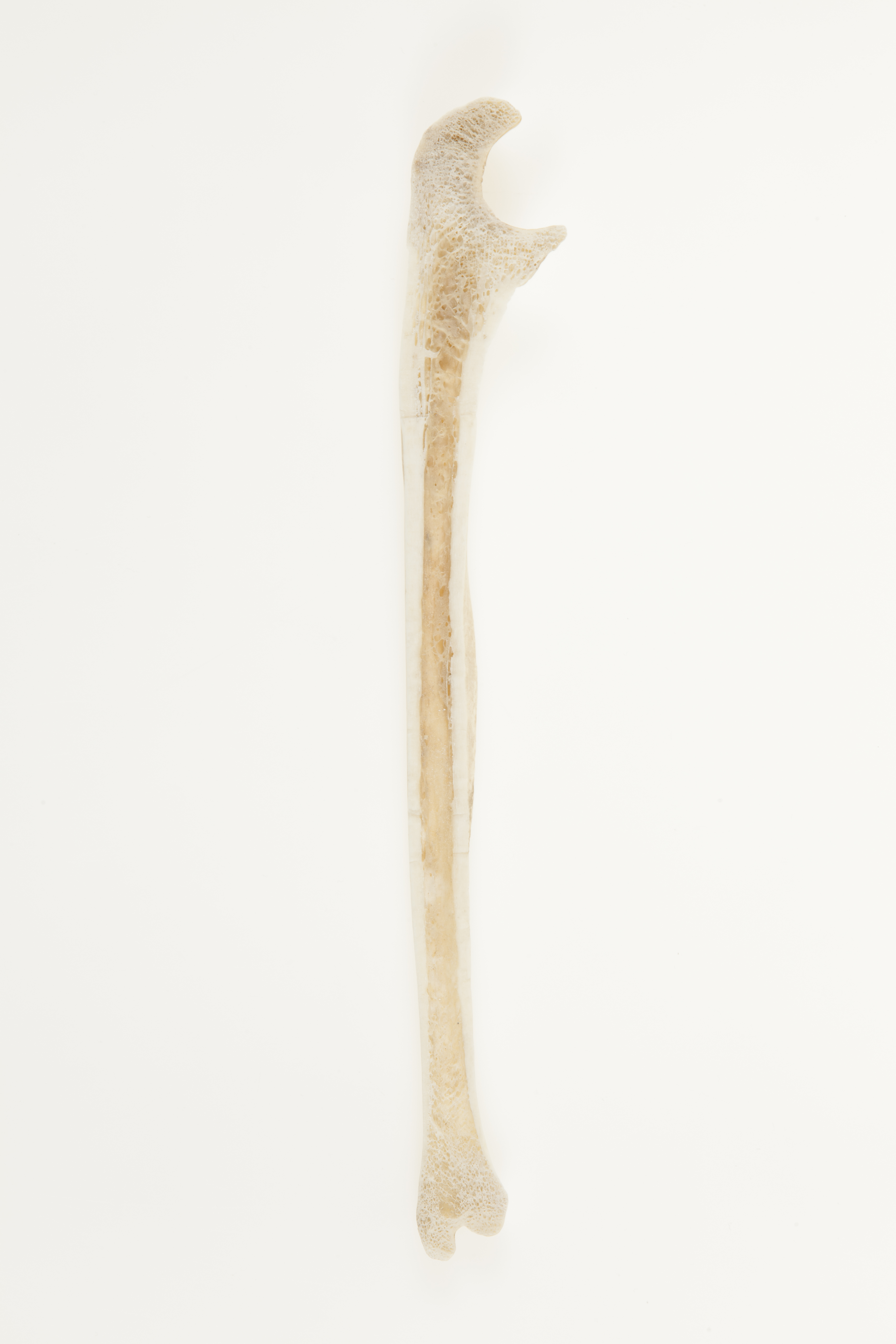
Ulna, coronal section
Adult ulna, section in coronal plane.
The medial bone of the forearm. The proximal shaft is triangular in cross-section. The distal quarter of the bone is round. In section, the compact (cortical) bone is seen around the shaft. In the adult long bone, the medullary cavity is where yellow marrow (a reserve of fat cells) is found. Spongy (trabecular) bone is seen in the end of long bones. In the growing skeleton, this is the site of red (blood forming) marrow.
Orientation: interior, lateral

|

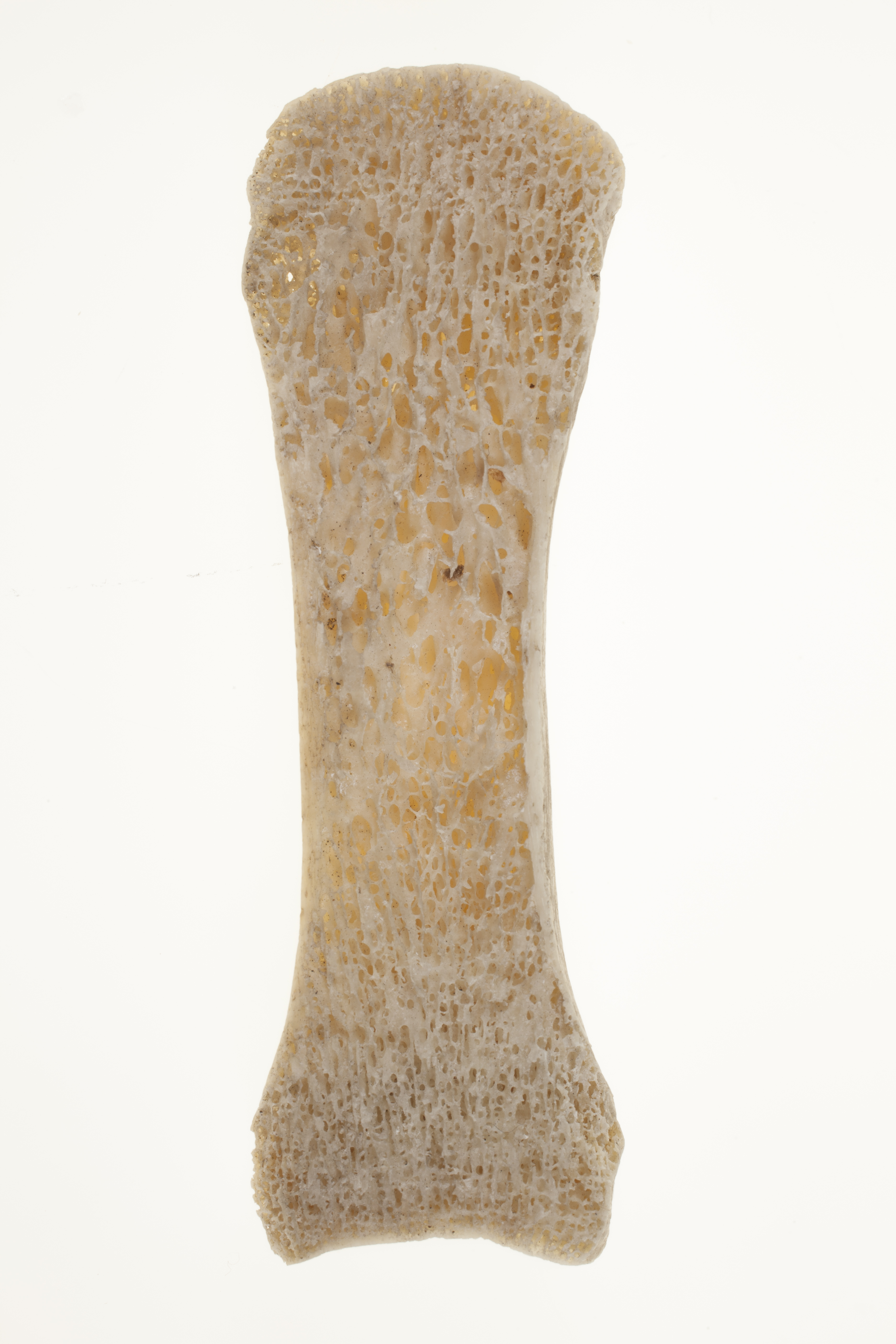
Metacarpal bone, coronal section
Adult first metacarpal bone, section in coronal plane.
The first metacarpal is the shortest and most robust of the metacarpals. The joint with the trapezium (the most lateral distal carpal bone) is where the majority of the movements of the first digit occur. The two gross structures of bone, compact (cortical) and spongy (trabecular), are seen in section.
Orientation: interior

|

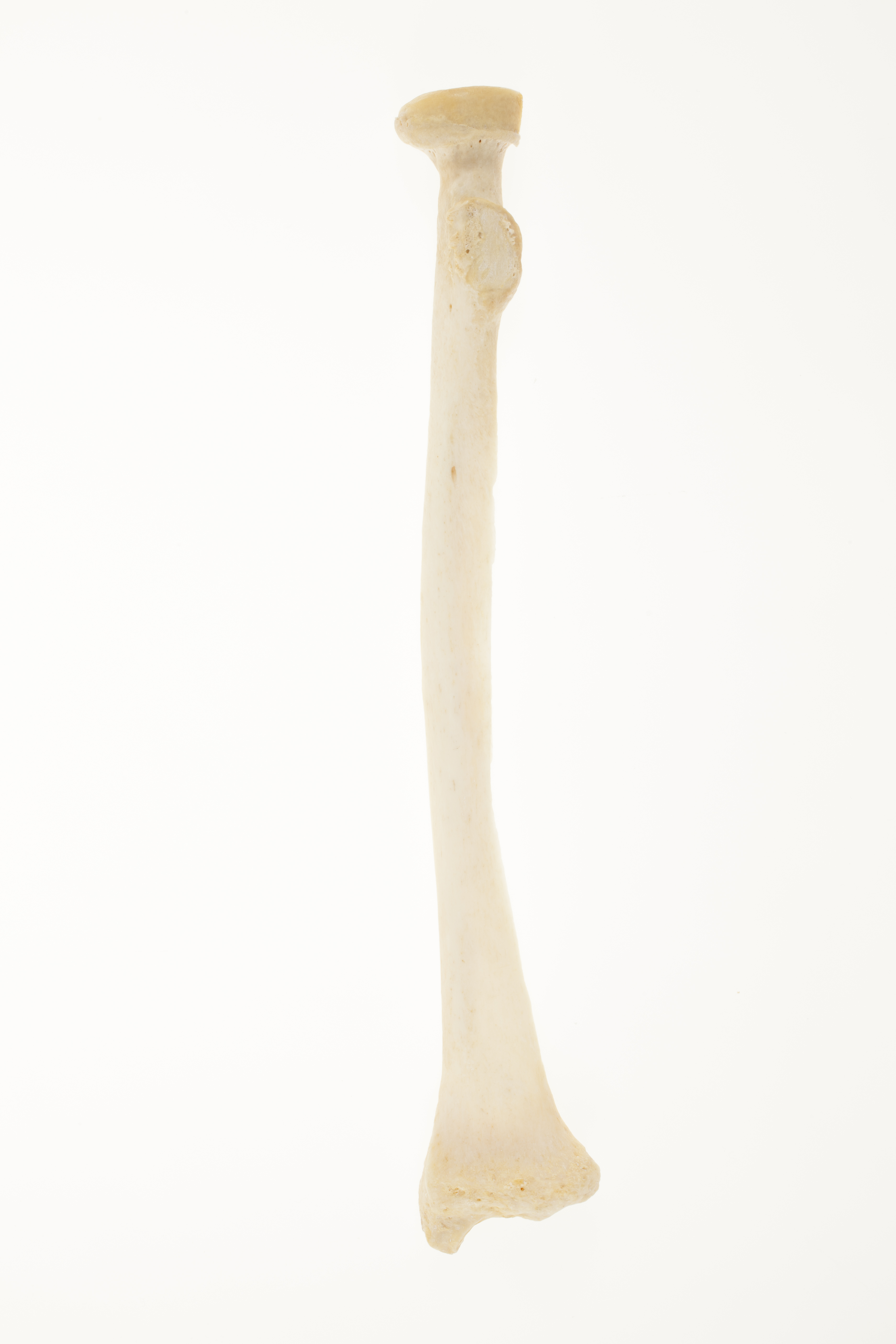
Right radius
Description: The radius is the lateral bone of the forearm. It moves relative to the ulna when we rotate our hand to face either palm up or palm down. When the palm is up, the radius and ulna lie parallel to one another. When the palm is down, the radius crosses over the ulna.
The head of the radius is cylinder-shaped, and articulates with a hollow in the ulna as well as with the capitulum of the humerus, as part of the elbow joint. The radial or bicipital tuberosity marks the insertion of the biceps brachii muscle, a flexor of the forearm. The oblique line spirals downwards and outwards from the tuberosity, and is the insertion point for muscles of the hand. The distal styloid process is an attachment point for the tendon of the brachioradialis (at the base of the process) and the radial collateral ligament of the wrist (at the apex). Distally, the radius articulates with two of the carpal bones of the wrist: the lunate and scaphoid.
Orientation: anterior

|
