The temporal bone has a unique place in anatomy. It contains two linear accelerometers, three angular accelerometers, a sophisticated organ of hearing, five cranial nerves, the jugular vein, and the carotid artery. If you climb, fly or dive, it is also a personal barometer. You will feel the urge to clear your ears every 300 feet in air, or every 5 meters in water. Contribution by Mr Roger Gray, Consultant Otolaryngologist, Cambridge Teaching Hospital and Fellow Murray Edwards College, Cambridge.
Dissected inner ear specimensThree mounted inner ear dissections, comprising one human specimen, and two ox specimens. The cochlea is concerned with hearing, whilst the semicircular canals, saccule and utricle are part of the vestibular system. MAN: note prominent semicircular canals and cochlear deeper in. Crus commune very clearly shown where the superior and poste rior semicircular canal share an entrance to the vestibule. OX 1: The specimen is mounted on a resin block. It displays a large diameter and rather thin semicircular canals, as well as a well delineated vestibular aqueduct and endolymphatic sac. OX 2: Showing semicircular canals and cochlea
|
|
|
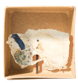
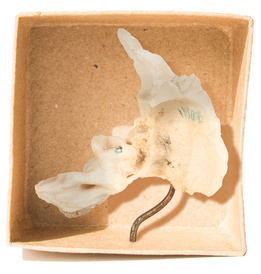
Disarticulated temporal boneDescription: Mastoid Process and squamous temporal bone sectioned to show extensive mastoid pneumatisation and sigmoid sinus (blue paint).
|
|
|
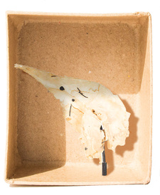
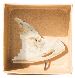
Disarticulated temporal boneDisarticulated squamous, petrous and tympanic parts to show styloid process, petrotympanic fissure and tympanic ring.
|
|
|
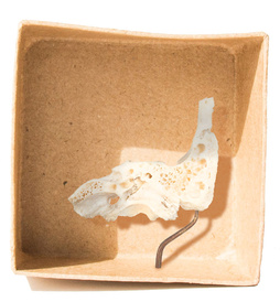
Disarticulated temporal boneComplete temporal bone with deep surfaces of mastoid and petrous removed to show extensive pneumatisation and relation of carotid canal in petrous apex.
|
|
Disarticulated temporal bone with burr holeBurr hole in squamous temporal bone to access pus and osteitis on posterior face of petrous temporal bone (otitic abscess in middle cranial fossa). Note sequestrum of bone in septic opening.
|
|
|
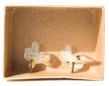
Dissected bony labyrinthDissection of bony labyrinth to show complete bony outline (on specimen on right) and passages within the labyrinth to show internal structure and site of the perilymph, membranous labyrinth, and endolymph (not visible) on specimen on left. Note round window and oval window on the specimen on the right.
|
|
|
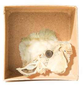
Petrous temporal bonePetrous temporal bone dissected to show bony labyrinth. Note the three semicircular canals at right angles to each other which detect movements of the head: pitch (rotation around side-to-side axis), roll (rotation around the front-to-back axis) and yaw (rotation round vertical axis). Note also the spiral of the cochlea.
|
|
|
|


