Department of Physiology, Development and Neuroscience
Thorax
Under construction
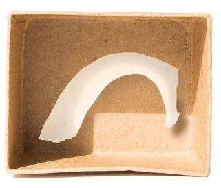
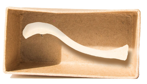
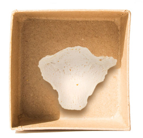
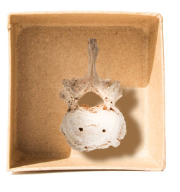
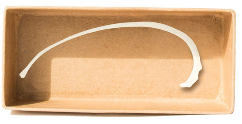
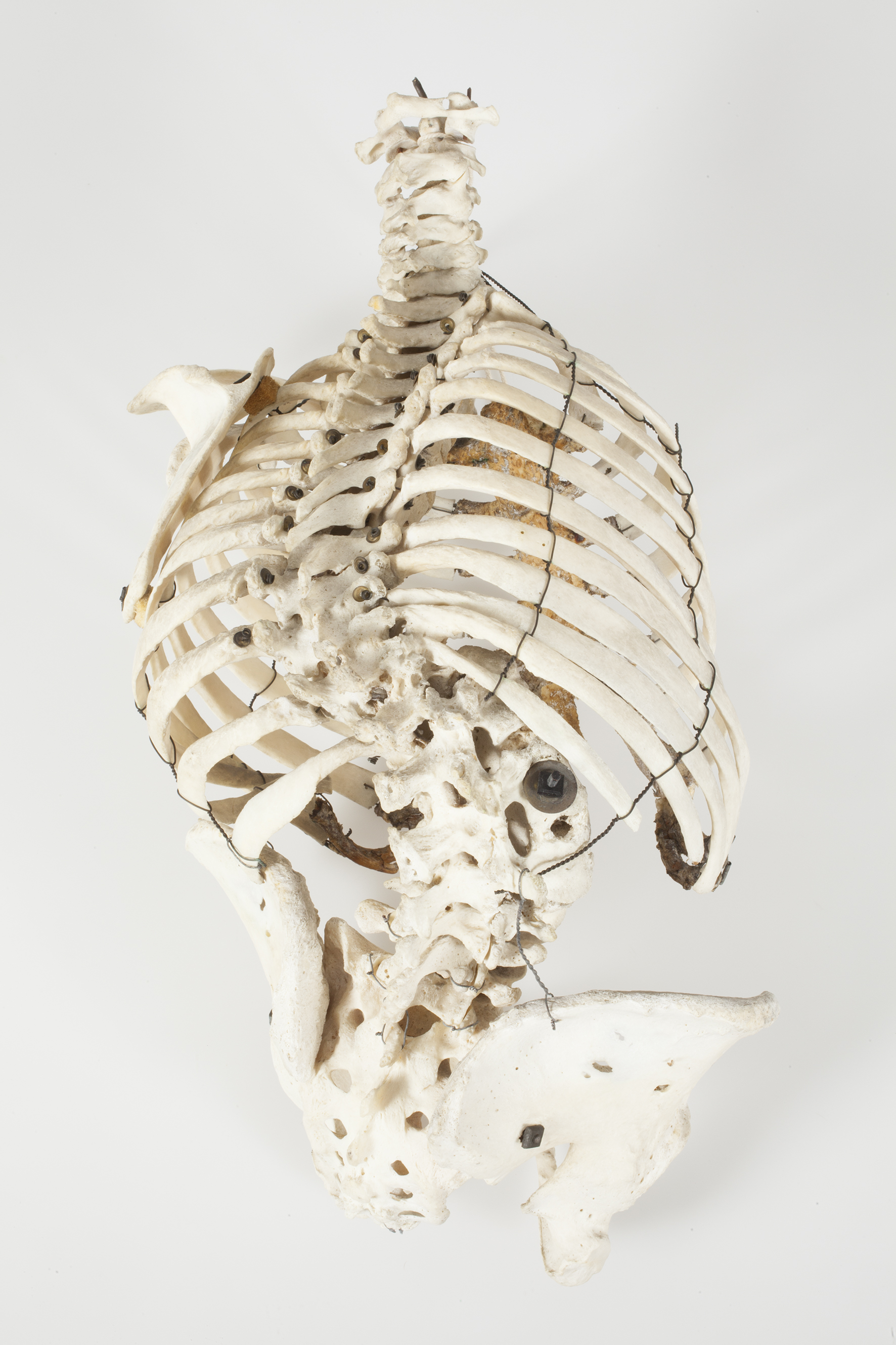
Sternum with calcified and deformed lower costal cartilages
Showing the manubrium, sternum, xiphisternum and attached costal cartilages with marked deformity of the xiphisternum and lower costal cartilages.
In the developing fetus, a ‘blueprint’ of the ribs is laid out in cartilage, which subsequently ossifies to form the rib bones. The costal cartilage is the persistent and non-ossified ventral extension of these cartilaginous models. The subsequent development of the costal cartage may be impacted by thoracic trauma, infection, ischemia, or by developmental or endocrine disorders. Calcification of the costal cartilage in adulthood is a normal process of aging.
Pathology: deformity of lower costal cartilage. Origin and pathology unknown.
Orientation: anterior
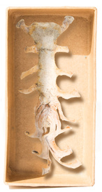
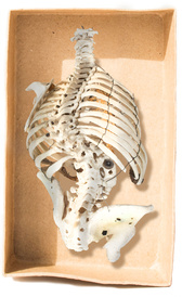
Posterior view of vertebral column demonstrating severe scoliosis
Scoliosis (Gr. “curvature”) is a lateral curvature of the vertebral (spinal) column. With the apex of the curvature sitting to the right of the midline and between the L2 and L4 lumbar vertebrae, this specimen can be described as showing a ‘right lateral lumbar scoliosis’. Also note the lateral deviation of the spinous processes, demonstrating that as scoliosis progresses the spine also begins to rotate on its longitudinal axis. The majority of scoliosis deformities are as a result of ‘Idiopathic Scoliosis’, however this condition can occur due to many other conditions, including neuromuscular disorders, spinal infections and following trauma. This severe example of scoliosis would have caused this patient significant disability in their day-to-day life. This not only occurs due to the skeletal deformity but also the resultant effects this has on the organs within the thoracic and abdominal cavities. This is evident from the asymmetrical rib cage (which would give the characteristic ‘rib hump’ on the right hand side when the patient was asked to bend forward) and abdominopelvic orientation (evident from the asymmetry between the right and left hemithoraces to their corresponding iliac crests and the significant pelvic tilt). In mild cases of scoliosis with no significant complications, bracing can be used to prevent further progression. In more severe cases or those with associated complications, surgical correction is indicated to correct the deformity.
Pathology: Scoliosis
Superior view of adult lumbar vertebra with alterations due to osteoarthritis
Mechanical stress on the vertebral column leads to several alterations of the osseous vertebral surface, visible on this specimen: the appearance of osteophytes (bony spikes) on the margins of the vertebral body, pitting of the body of the vertebra, and alterations of the facet joints. In this case, there are also present destructive lesions of the centrum. It is likely that the individual suffered from lower back pain. The specimen displays post-mortem damage to the transverse processes and articular facets (partly broken). Furthermore, two round holes (for the passing of strings that would have held several vertebrae together) puncture the body of the specimen, reminders of the use of this vertebra for anatomical teaching.