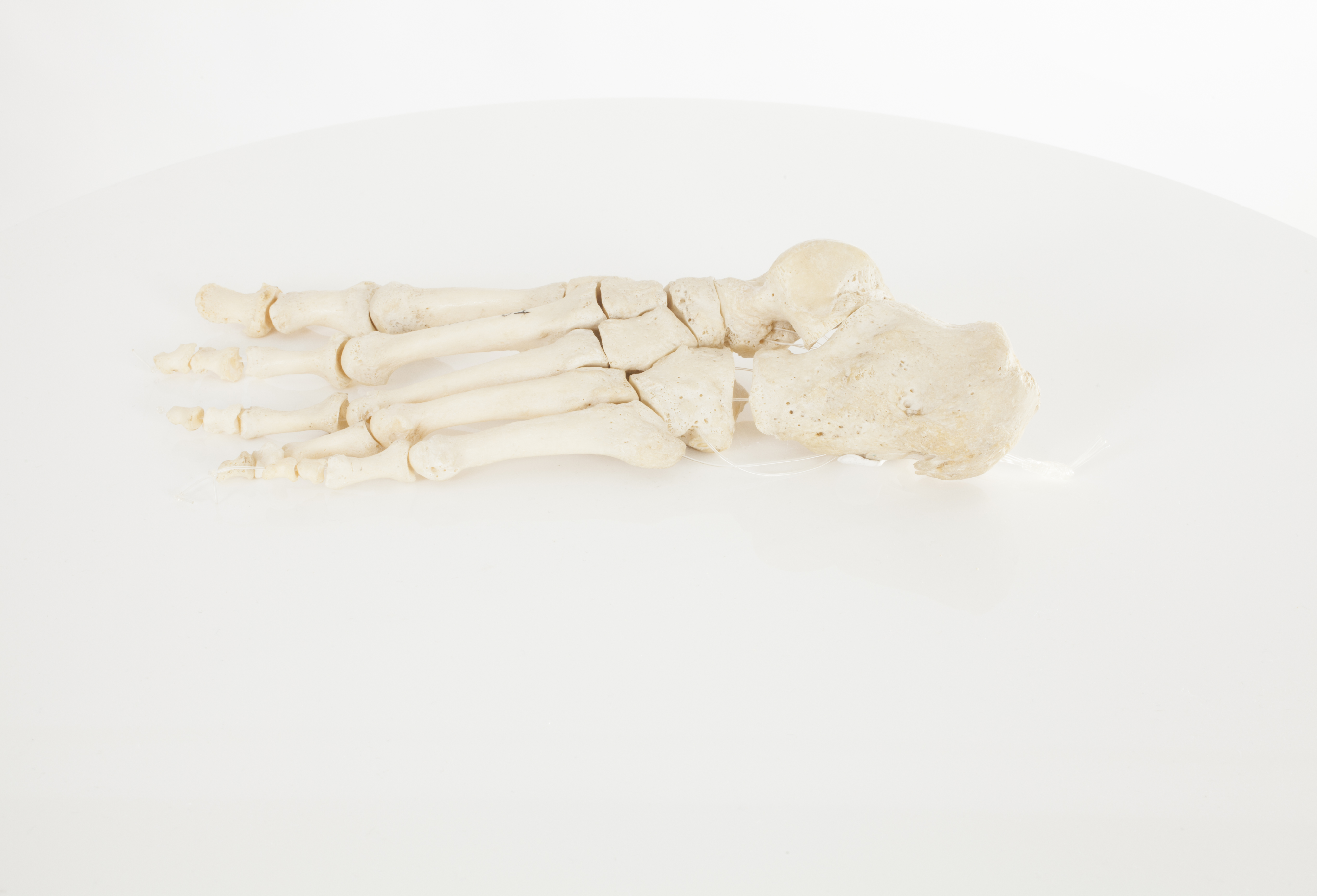
 Foot Foot
Description: Adult foot. The human foot is composed of 26 bones, grouped in three segments: tarsals, metatarsals and phalanges. On the calcaneus, commonly referred to as the heel bone, is where the Achilles tendon attaches, the thickest tendon in the human body.
Pathology: Enthesopathy on the calcaneus
 
|
|
|
|
|
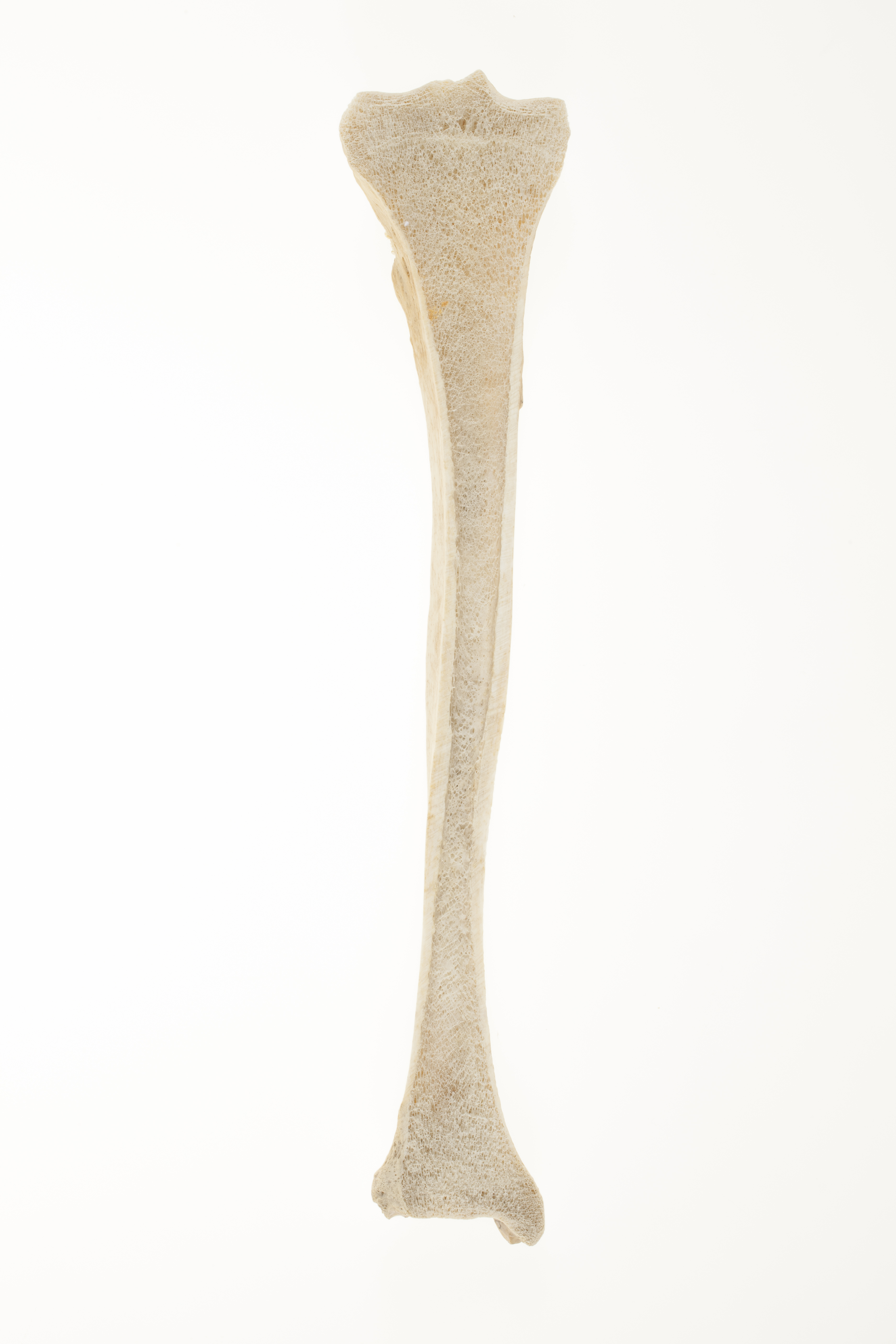 Tibia, coronal section (internal) Tibia, coronal section (internal)
Description: The tibia is the larger and stronger of the two bones below the knee. It is the main weight-bearing bone of the lower limb and provides attachment to muscles that move the foot. It articulates with the femur to form the knee (proximally), and with the talus to form the ankle (distally). The main features at the proximal end are the tibial plateau, with the projecting intercondylar eminence, which separates the two articular surfaces. On these sit the menisci, which are fibro-cartilaginous, crescent-shaped discs that cushion the knee joint.
At the distal end, the medial malleolus projects downwards. The word malleolus is from Latin, and means ‘a little hammer’. The malleolus ends in a thin groove, which is the attachment point for the capsule of the ankle joint.
In a growing child, the tibia is comprised of three main sections: the shaft or diaphysis, and, at either end, the proximal and distal epiphyses. About the age of 13-19 years, the ends fuse on to the main shaft to create one bone. The line of fusion - the epiphyseal line - can be seen here in the sectional view of the tibia, at the proximal end.
Orientation: anterior
 
|
|
|
|
|
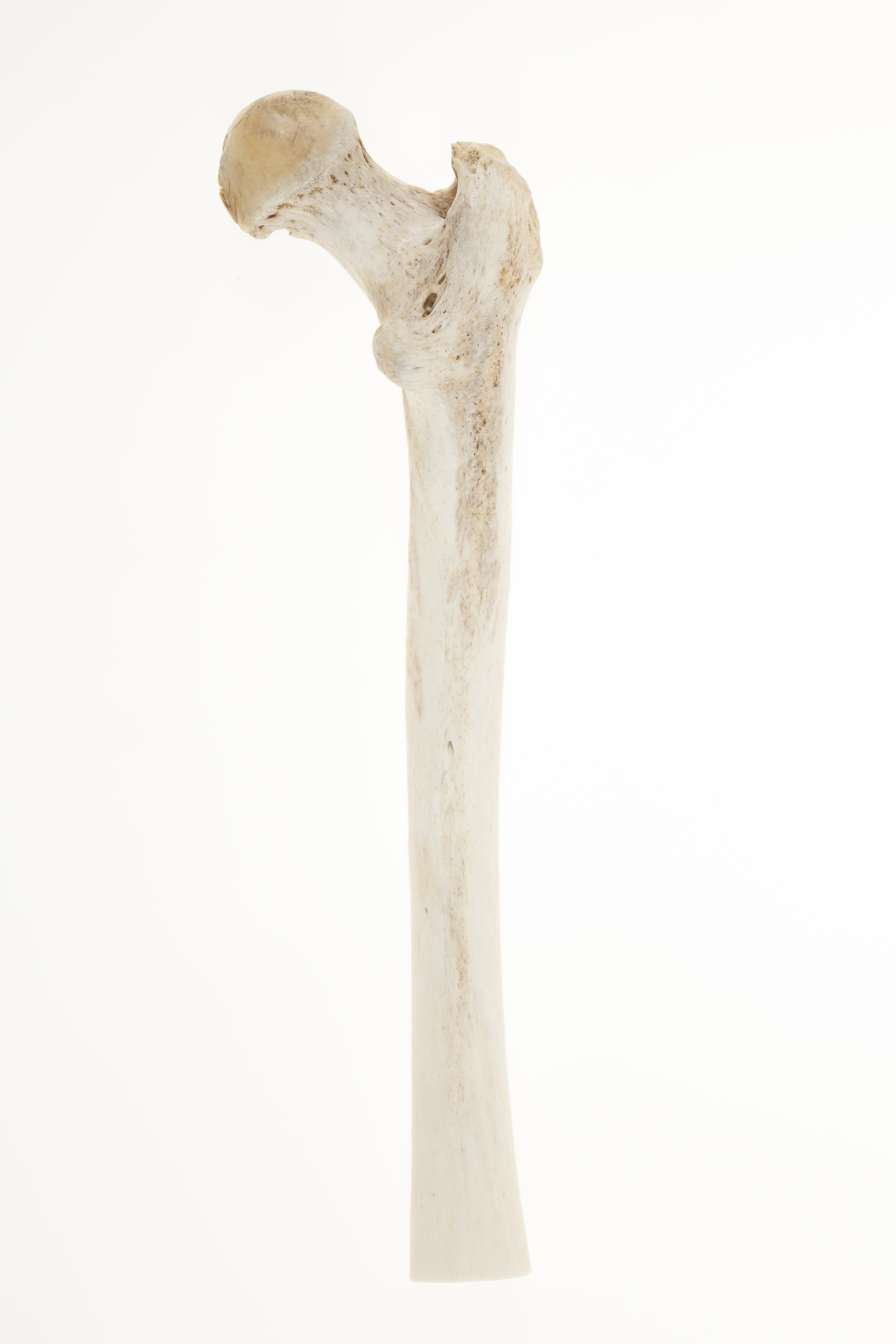
Right partial femur: posterior
The shaft of the femur is under some degree of torsion, that is, it is twisted on its long axis. This is a normal result of the difference in angle between the head and the distal end. The degree of torsion may be related to activity levels, and might influence the mechanics of the hip.
In the middle third of the posterior surface can be seen the linea aspera, which is a roughened area where muscles attach. Specifically, to the inner side, insert the vastus medialis, adductors longus and magnus muscles, and the medial intermuscular septum, which is a fold of deep fascia (a sheet of fibrous tissue enclosing a muscle group). To the outer side attach the vasti lateralis and intermedius muscles, the short head of biceps femoris, and the lateral intermuscular septum.
The blood supply of the femur is profuse (plentiful). Fractures can lead to significant and dangerous blood loss. The arteries of the shaft run along the linea aspera, and enter via a nutrient foramen.

|
|
|
|
|
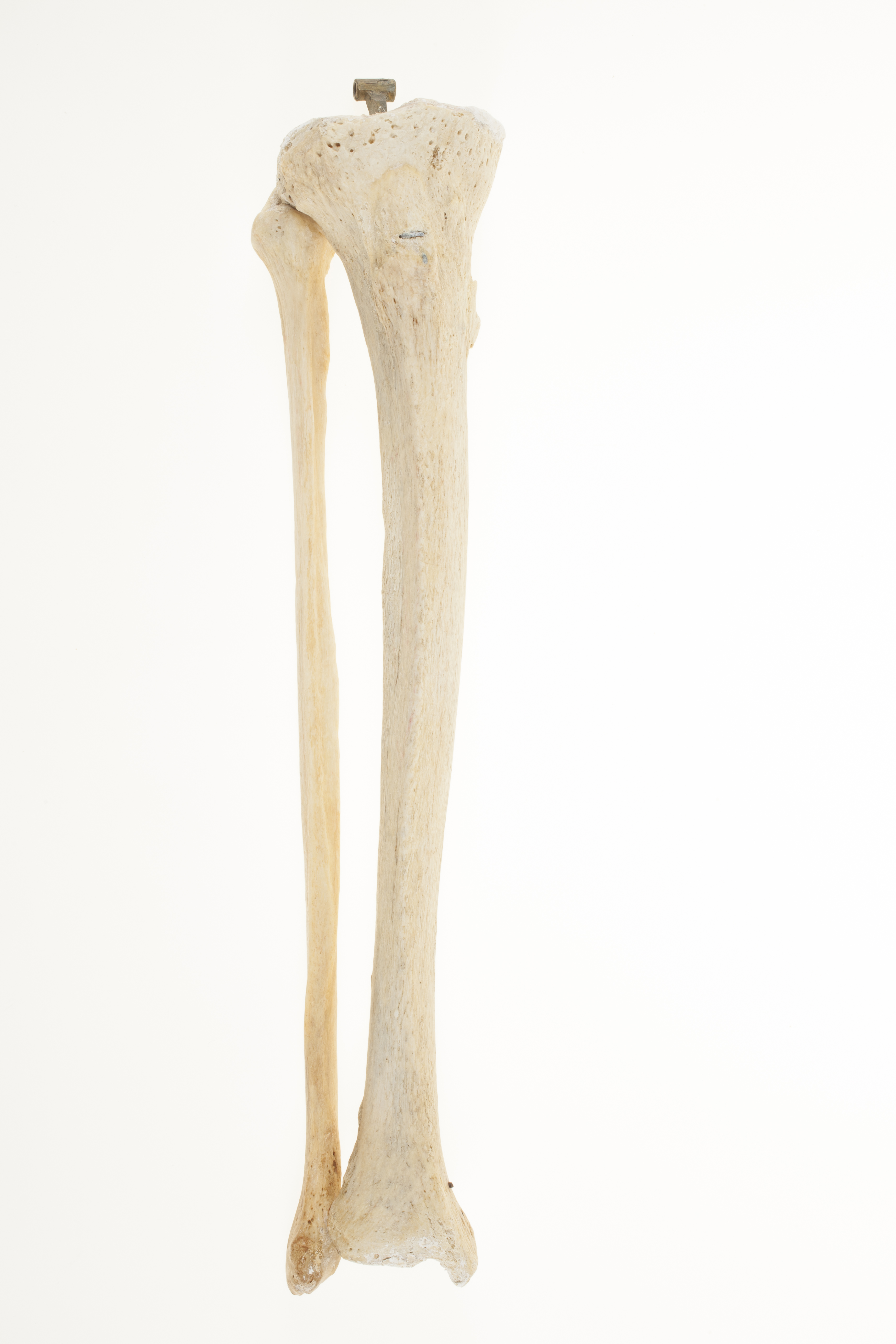
Right articulated tibia and fibula
Adult tibia and fibula. The two long bones of the lower leg play multiple roles: while the tibia contributes to supporting the weight of the body, the fibula helps in stabilizing the ankle joint. The name fibula derives from the Latin word designating a brooch for fasting clothes, as its position lateral to the tibia is redolent of a safety pin. The pair of bones are connected through metal rods (one being visible beneath the tibial tuberosity). There is slight damage to the distal epiphyses.

|
|
|
|
|
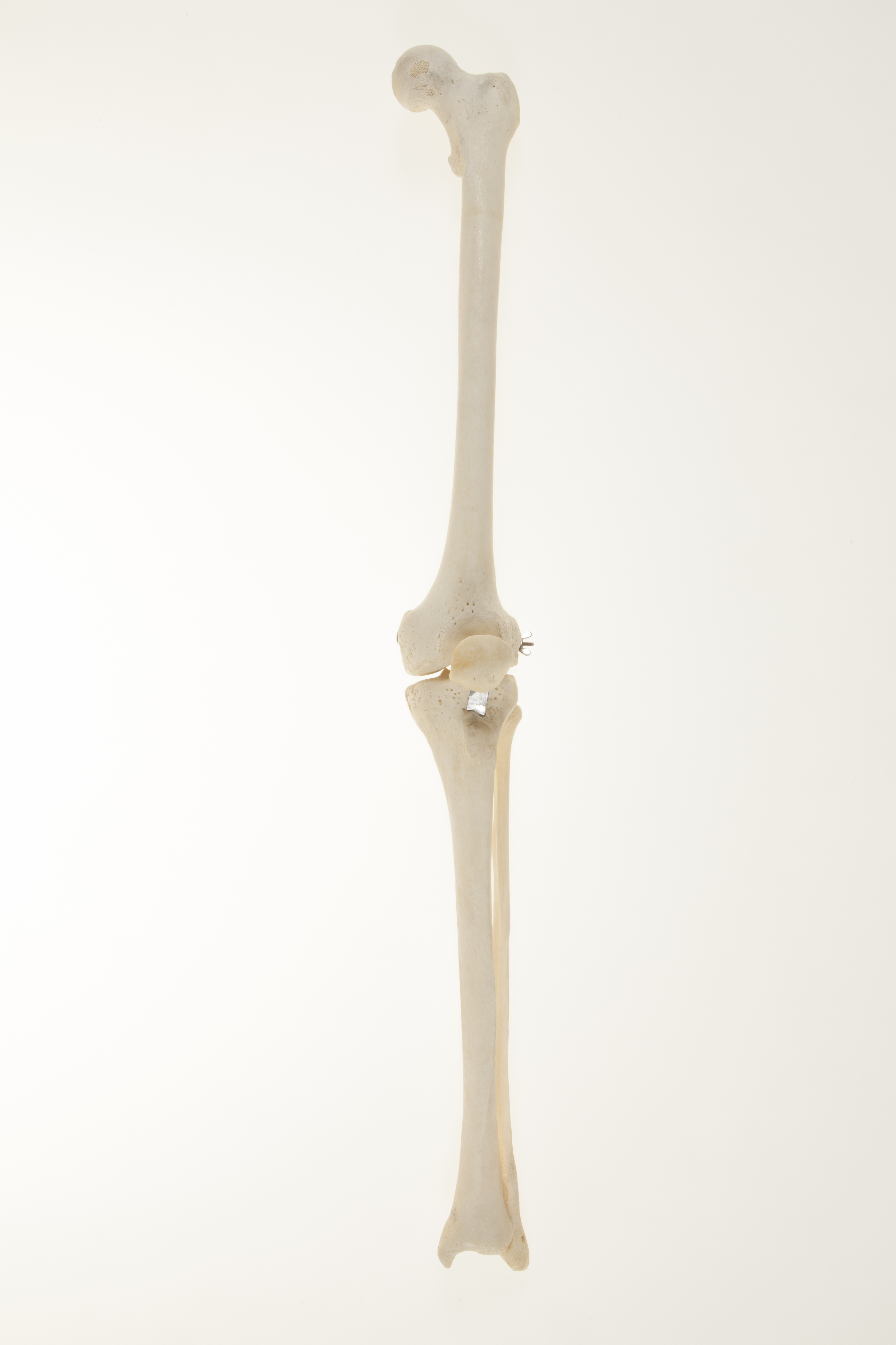
Left articulated femur, tibia, fibula, patella
The articulated long bones of the left adult lower limb, with patella at the knee joint.
The robust bones of the lower limb support the weight of the upper body, facilitate locomotion, and contain strong, stable joints.
The head of the femur (the thigh bone) articulates with the pelvis, thus forming the hip joint; the patella (or kneecap) articulates with the femur, and together with the
tibia form the knee joint; the distal end of the tibia and fibula form part of the ankle joint.
Orientation: anterior

|
|
|
|
|
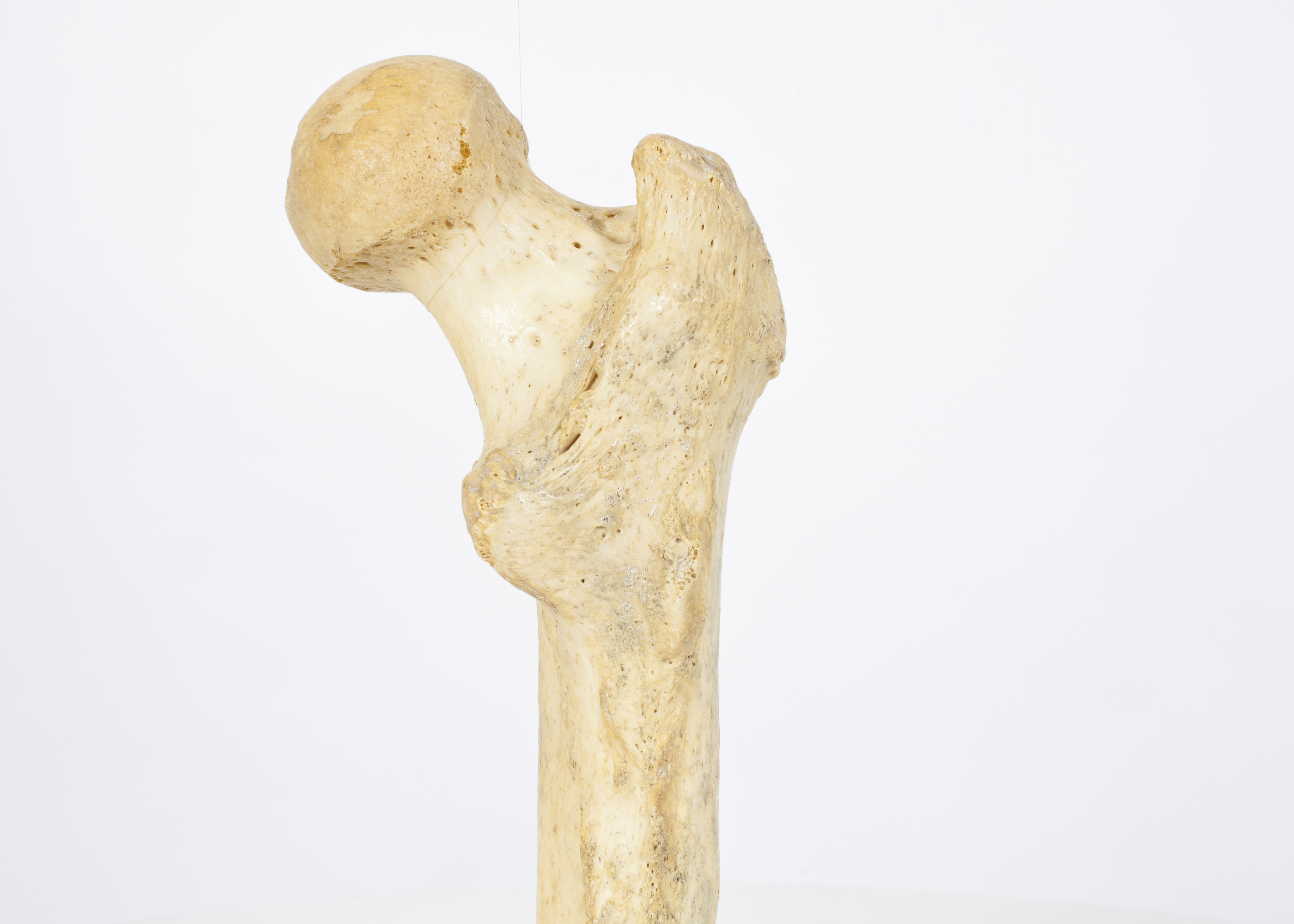 Right proximal femur Right proximal femur
The femur is the longest, strongest and heaviest of the bones in the human body. The femoral neck forms an angle of about 125° with the shaft of the femur.
The proximal end of the femur carries the greater and lesser trochanters. The greater trochanter is the insertion point of gluteus minimus and gluteus medius muscles, which help to abduct (move away) and medially (inwardly) rotate the thigh at the hip. The lesser trochanter is the insertion of the iliopsoas tendon, iliacus muscle and psoas major muscles which flex the thigh at the hip. The head of the femur fits into the acetabulum of innominate bone of the pelvis.
Orientation: posterior
 
|
|
|
|
|
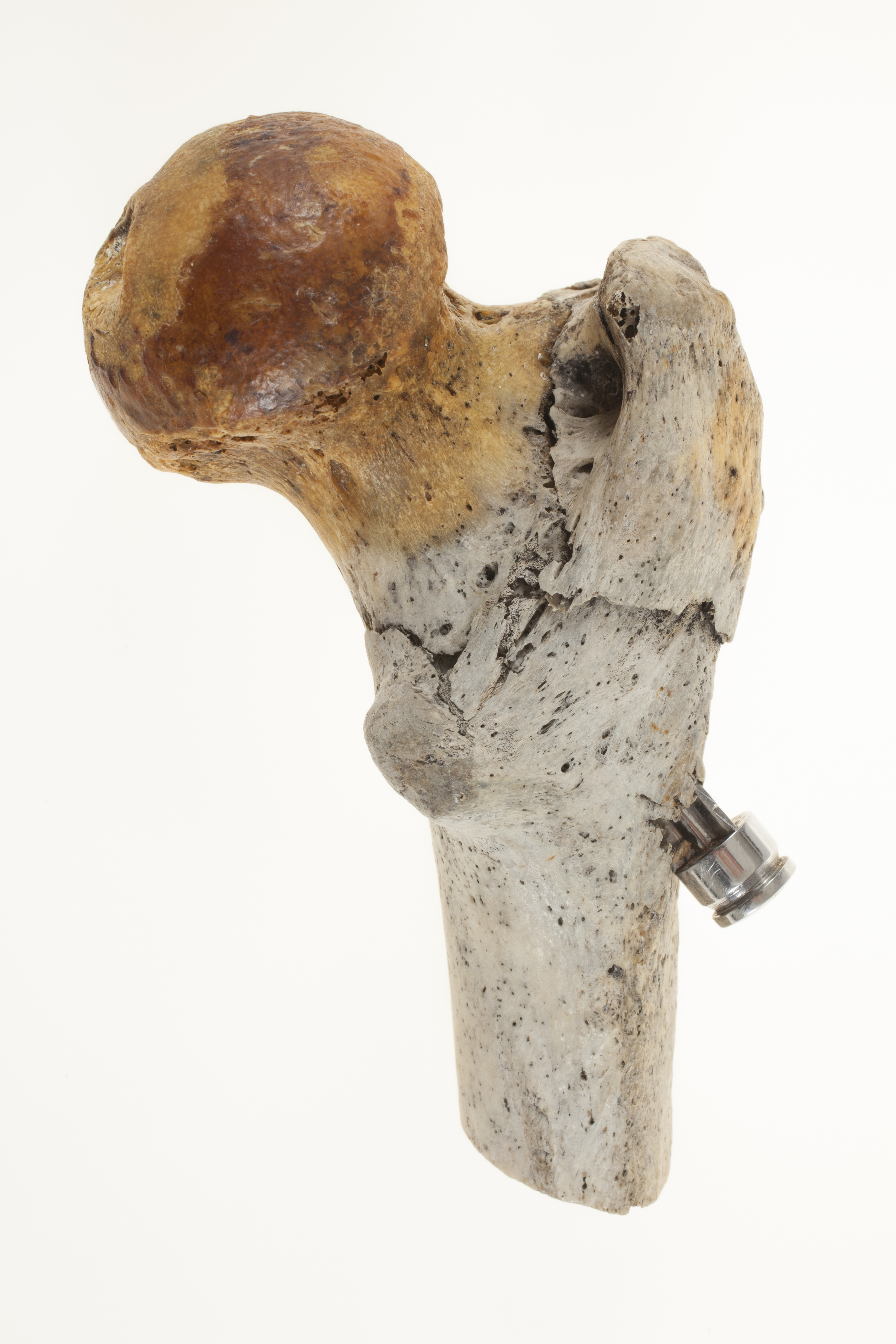
Right proximal femur, extracapsular neck of femur fracture, with dynamic hip screw in situ
Proximal section of right femur showing an old intertrochanteric fracture of neck of femur which has been operatively repaired using a dynamic hip screw. The femoral neck is the most common site for fracture of the leg in older women.
Pathology: fractured neck of femur
Orientation: posterior

|
|
|
|
|
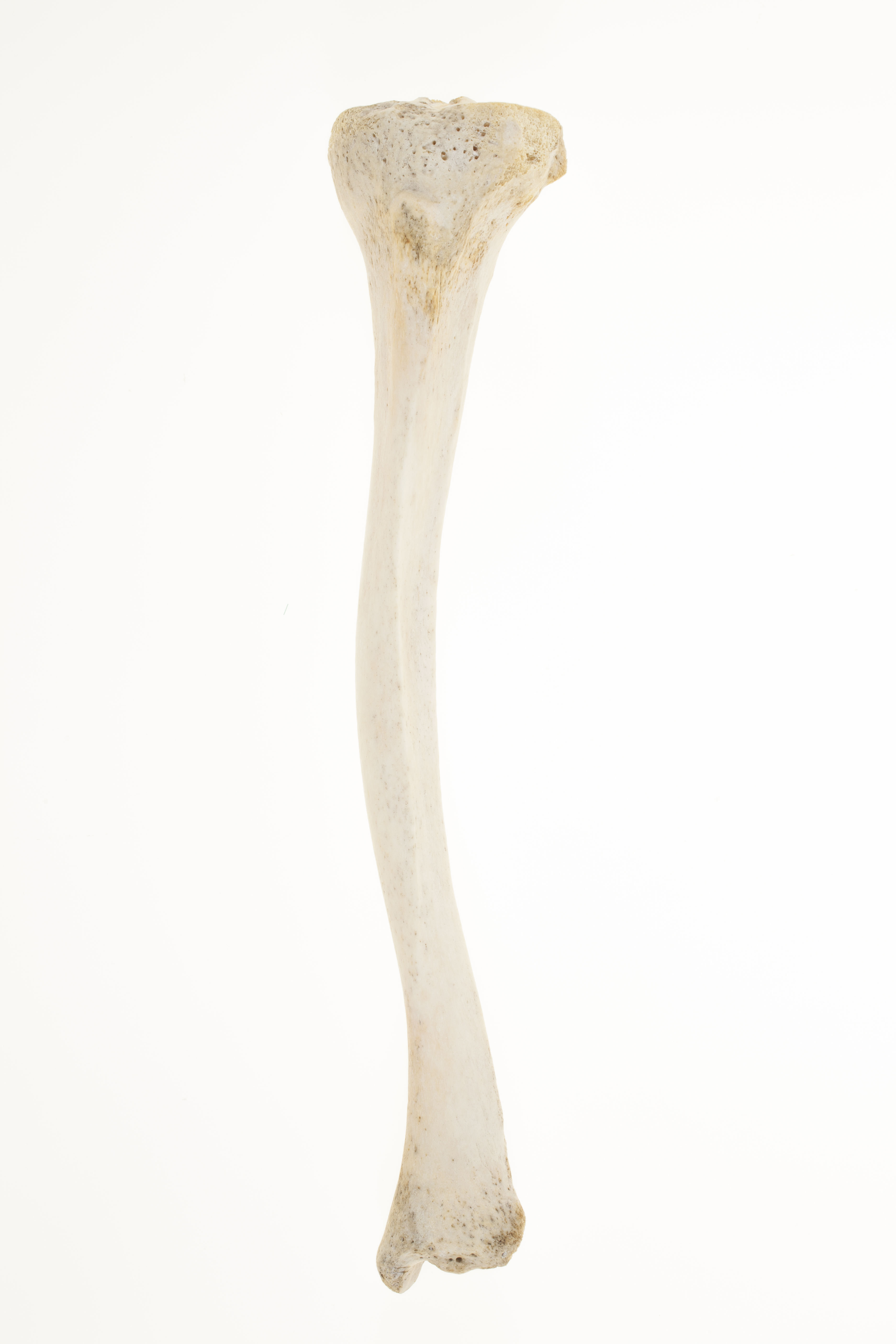
Adult tibia, showing bowing effects due to rickets
Vitamin D chronic deficiency during a child’s developmental phases leads to the deformation (inward bending) of the weight-bearing bones of the legs, as well as the expansion of the extremities of these bones, which can take a trumpet like shape . In living patients, lower extremity bowing can be due to several others factors, ranging from developmental bowing, to tibia vara (Blount disease) or achondroplasia, and clinical diagnosis alongside x-rays can differentiate between the diagnoses.
Pathology: Rickets
Orientation: anterior
|
|
|
|
|
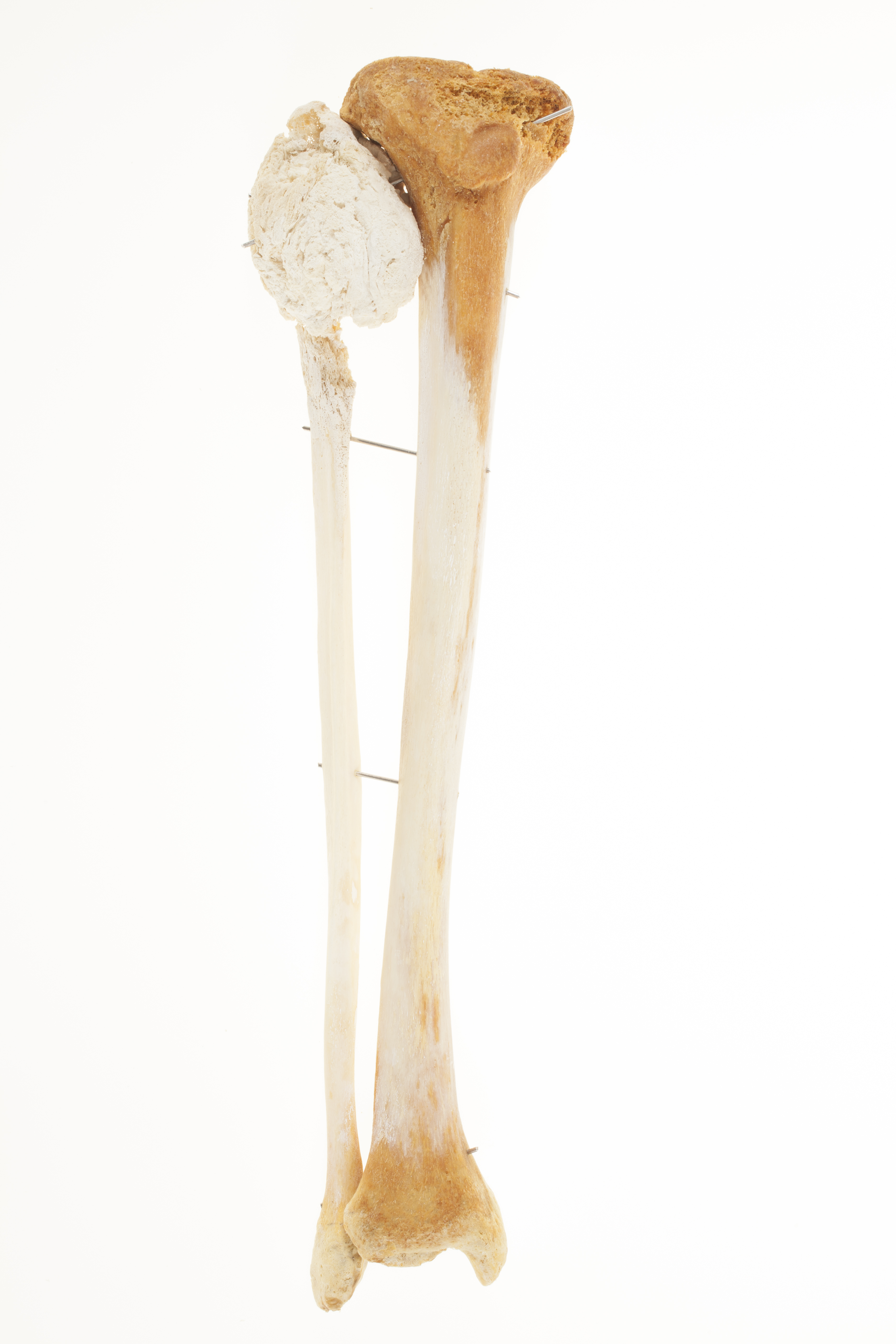
Tibia and fibula, with neoplasm
Right articulated adult tibia and fibula
The epiphysis of the fibula shows erosion of the cortical bone, and a large mass of expansive calcification. The contours of the fibular head are completely destroyed. Possible chondrosarcoma.
Pathology: neoplasm
Orientation: anterior

|
|
|
|
|
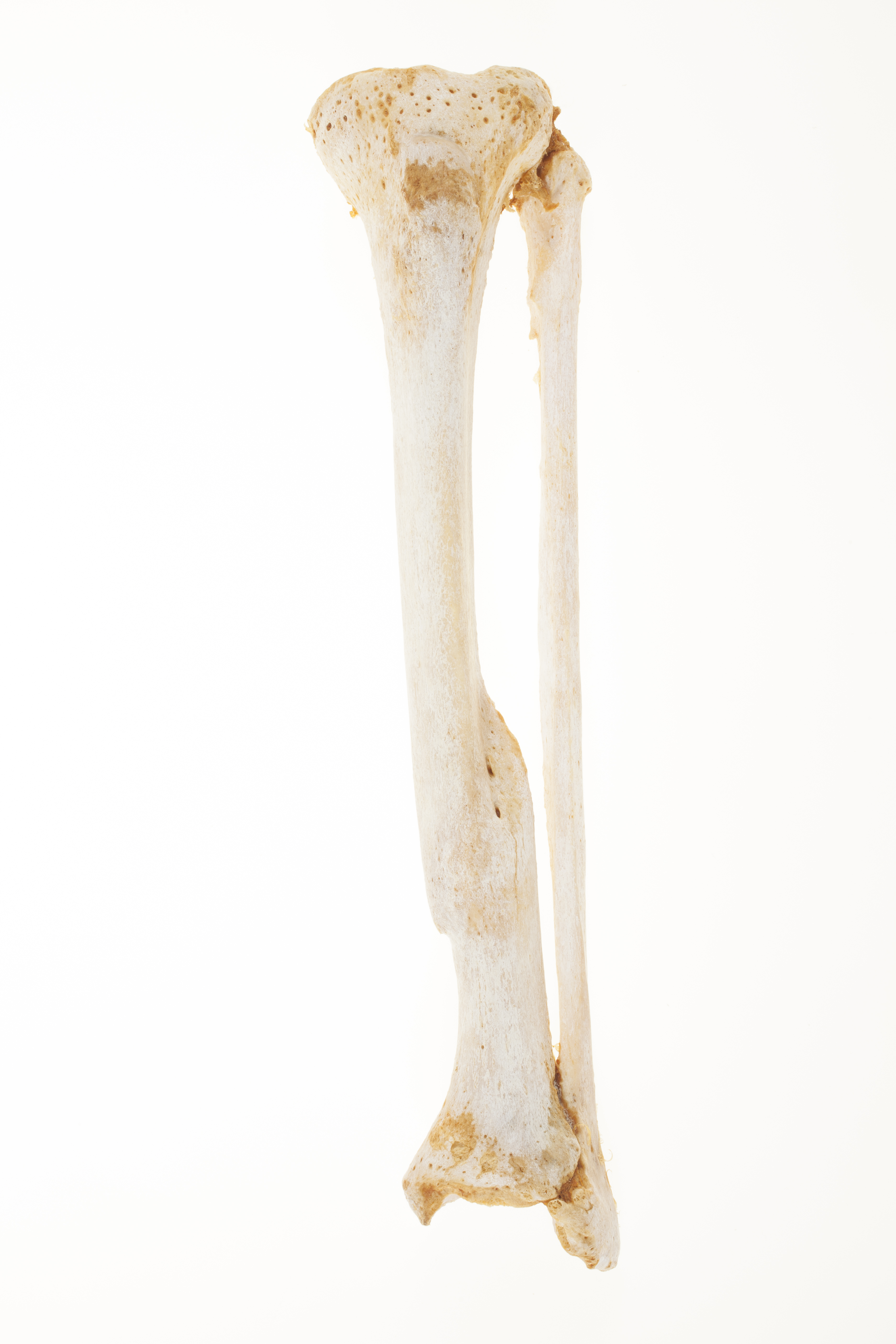
Left articulated fractured tibia and fibula
Anterior view of an adult fractured tibia and corresponding fibula. The tibia displays an oblique healed fracture in the distal third segment, with angulation and callus (the new tissue formed during the healing process around the margins of the broken bone). The process of bone remodelling in an adult can take several months. The two bones are articulated through the connecting ligaments.
Orientation: anterior
Pathology: Fracture
|
|
|
|
|
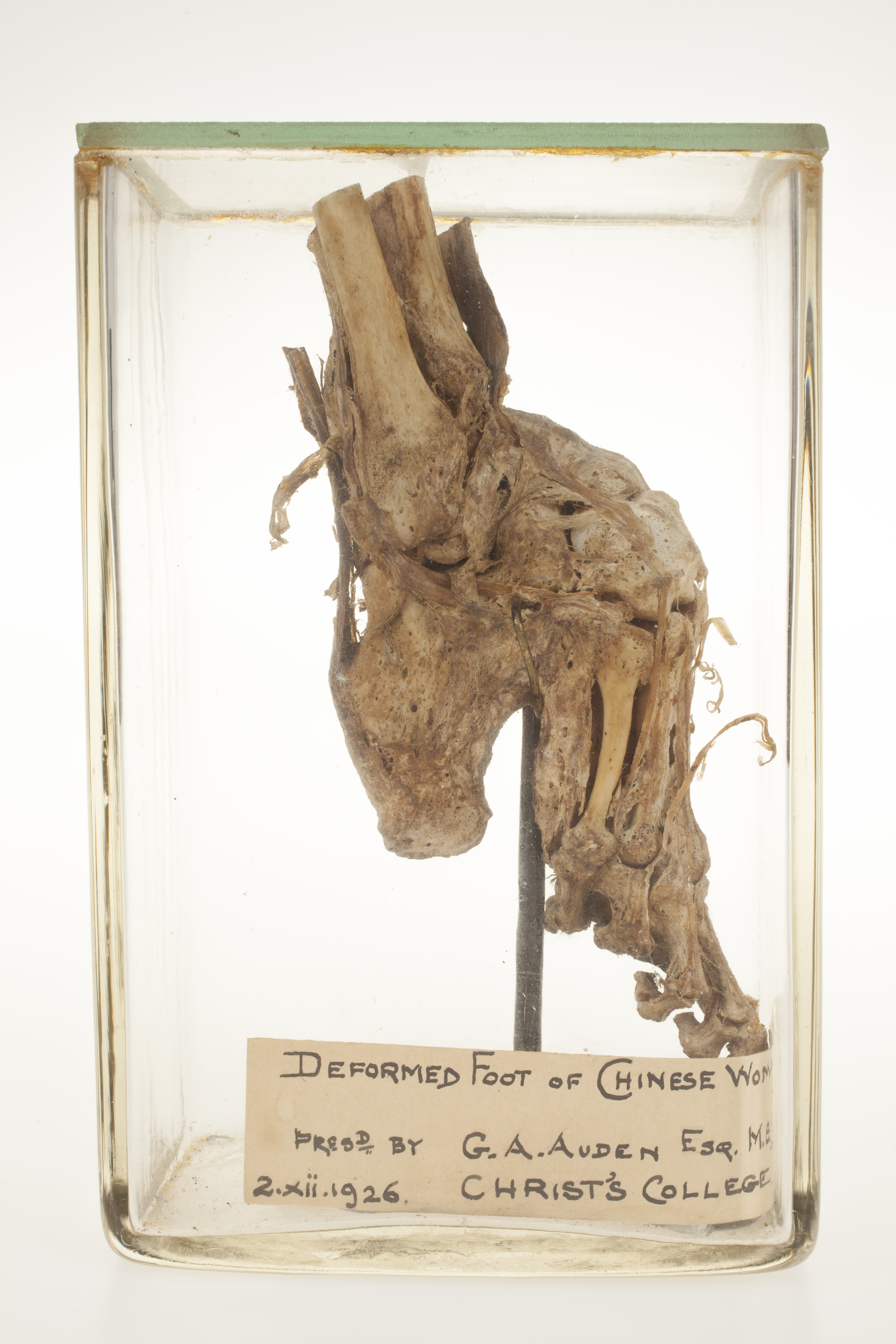
Bound foot of an adult woman
Skeletal specimen of an adult deformed foot which shows the effects of foot binding. The foot and the lower part of the leg bones (fibula and tibia) are present. The ligaments and tendons are also preserved. The practice of foot binding started in China during the Middle Ages, and consisted of breaking the toes of usually young girl’s feet, pressing them in the sole of the foot, also forcing the fracture of the arch of the foot, then tightly binding the foot, with the desired aim of shortening its length. These were called ‘lotus feet’, and became a symbol of status and beauty. The practice was formally banned in 1912. This specimen allows the observation of the effects of the procedure- the very acute angle between the metatarsals and calcaneus, as well as the inward folding of the toes, under the sole of the foot. The foot is accompanied by the label: ‘Deformed foot of Chinese woman. Presented by G.A. Auden Esq. M. B. December 2nd 1926. Christ’s College’.
Pathology: Bound foot
Orientation: Lateral |
|
