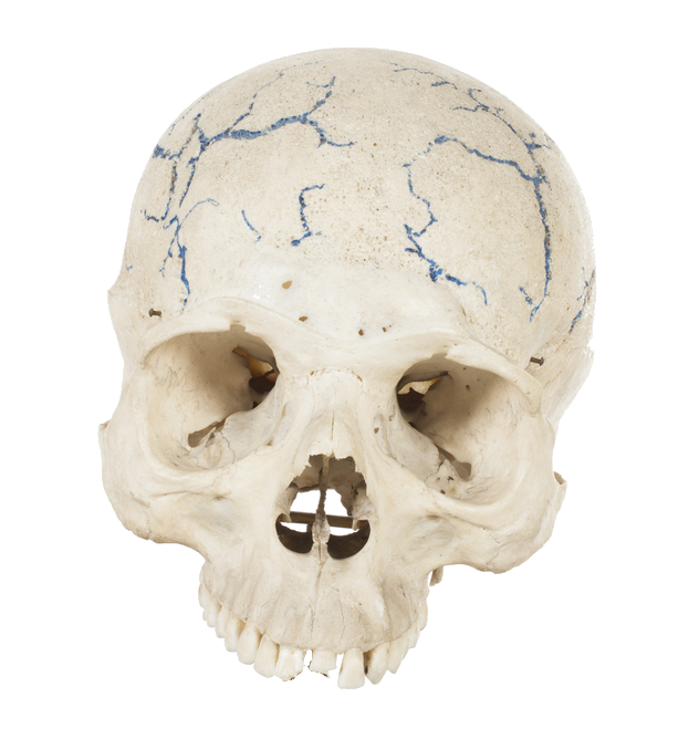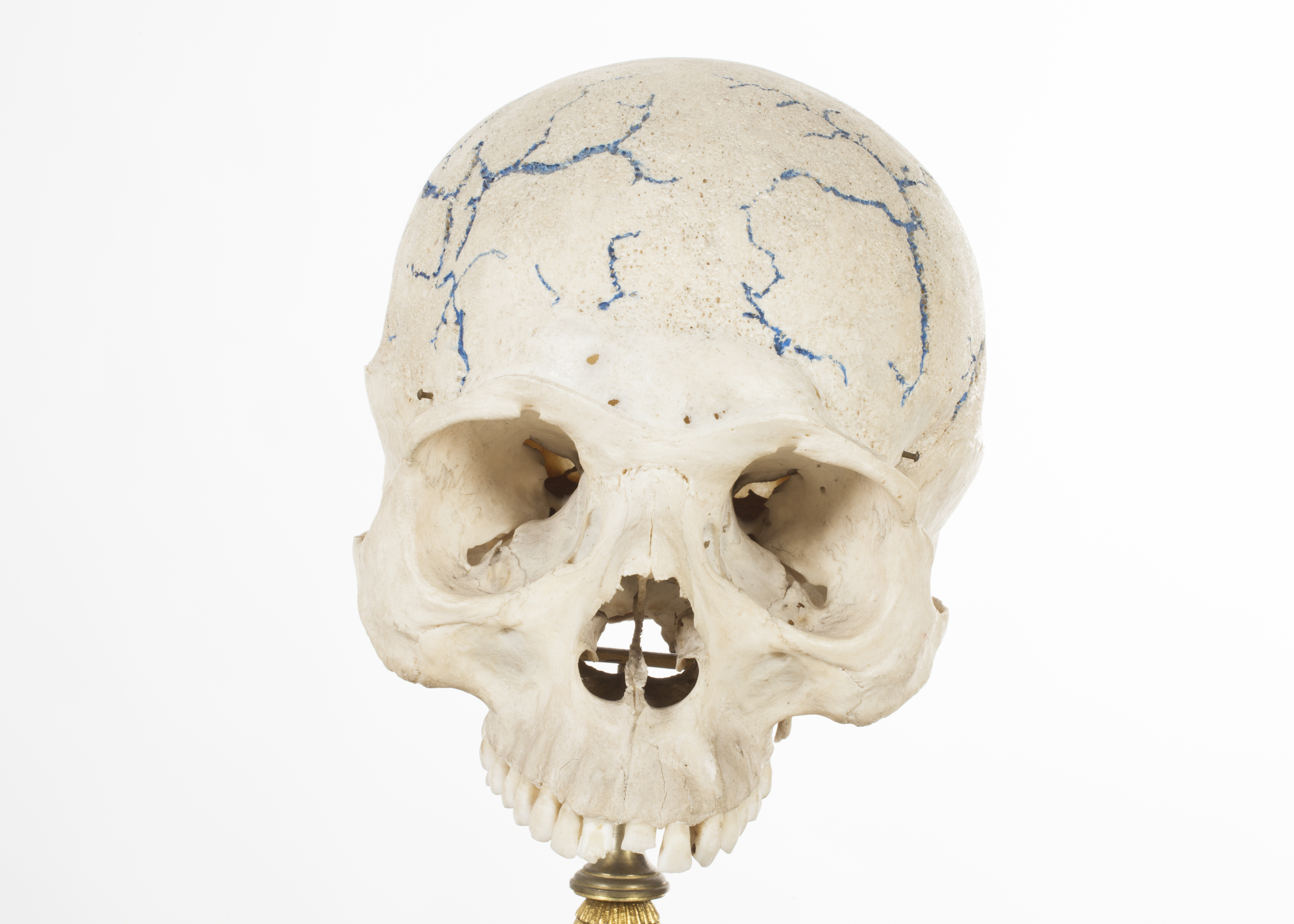
Skull with marking of scalp vessels
Description: Adult skull on to which have been added in blue paint the superficial blood vessels.

|
|
|
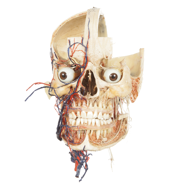
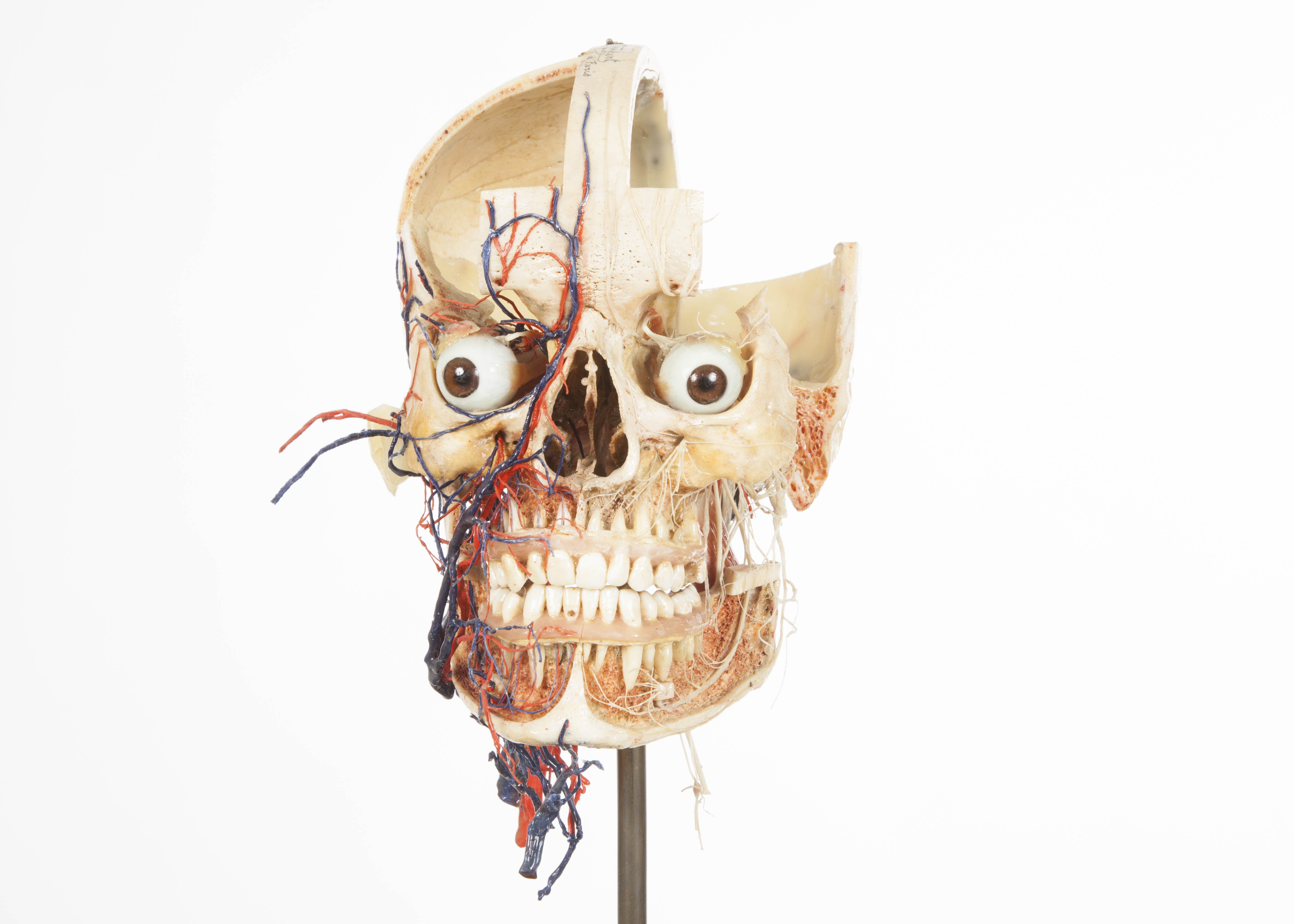 Dissected skull demonstrating the blood supply to the head and neck. Bone with wax. Mid to late 19th century. Maison Tramond. Dissected skull demonstrating the blood supply to the head and neck. Bone with wax. Mid to late 19th century. Maison Tramond.
Adult skull. The cortical bone on the maxillae and mandible has been removed in order to expose the dental roots, the blood supply and innervation system, as well as the spongy bone (here shown in pink). The infraorbital nerves are also visible, as well as the blood supply to the face on the right side. The blood supply to the head and neck is a complex system, the face being a richly vascularised area. The gums are modelled out of pink wax. The frontal squama (except the median area) and the left parietal bone have also been removed in order to expose the internal sagittal sulcus and meningeal grooves- the bone structures which house the blood vessels that supply/drain blood from the brain. The specimen is labelled in blue ink along the sagittal suture: ‘N Rouppert a Paris’. 
|
|
|
|
|
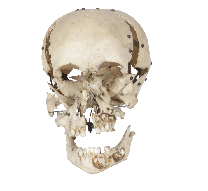
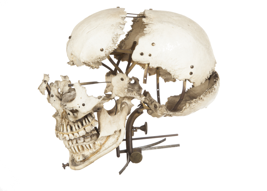
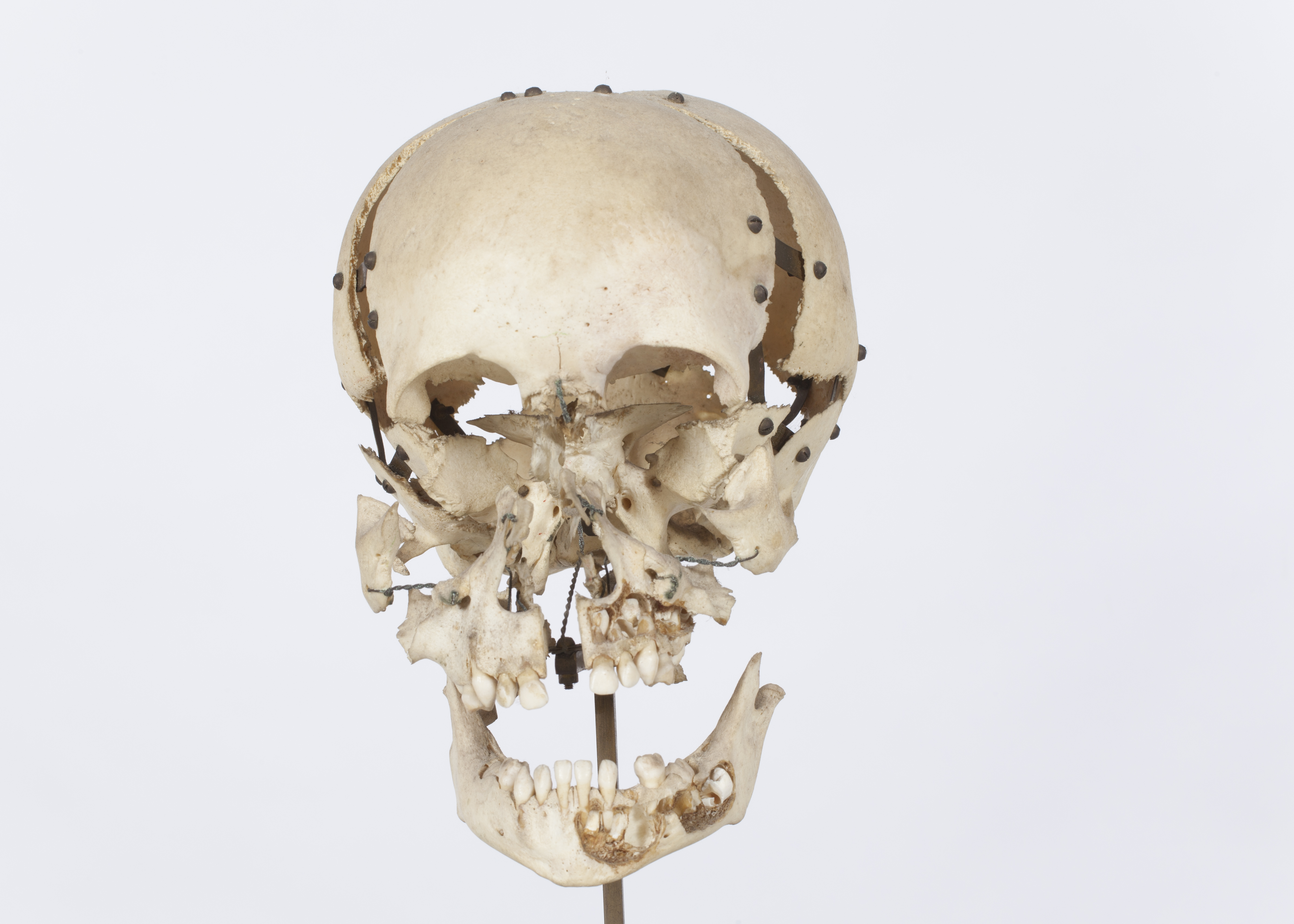 Beauchene skull Beauchene skull
Bone with brass connections and wires. Mid to late 19th century. Maison Tramond.
Child skull (with label marked in ink '1 yr 18mo'). Skull separated on the suture lines and joints in 13 parts, demonstrating the anatomical relations between the cranial and facial bones. The cortical bone layers on the left side of the mandible are cut out in order to expose the permanent dentition. In every child’s jaw the permanent dentition is present, slowly forming and waiting to erupt (starting around 7 years old). The dentition is a good indicator for age estimation, and is used for this purpose by forensic anthropologists and osteologists. The specimen is in almost perfect condition, with minor stains from the metal wires.

|
|
|
|
|
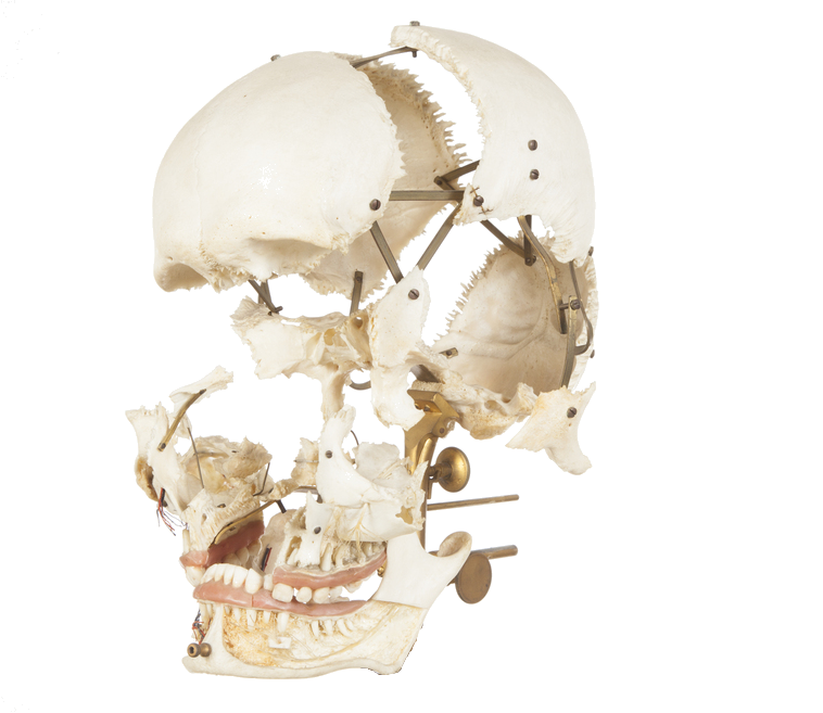
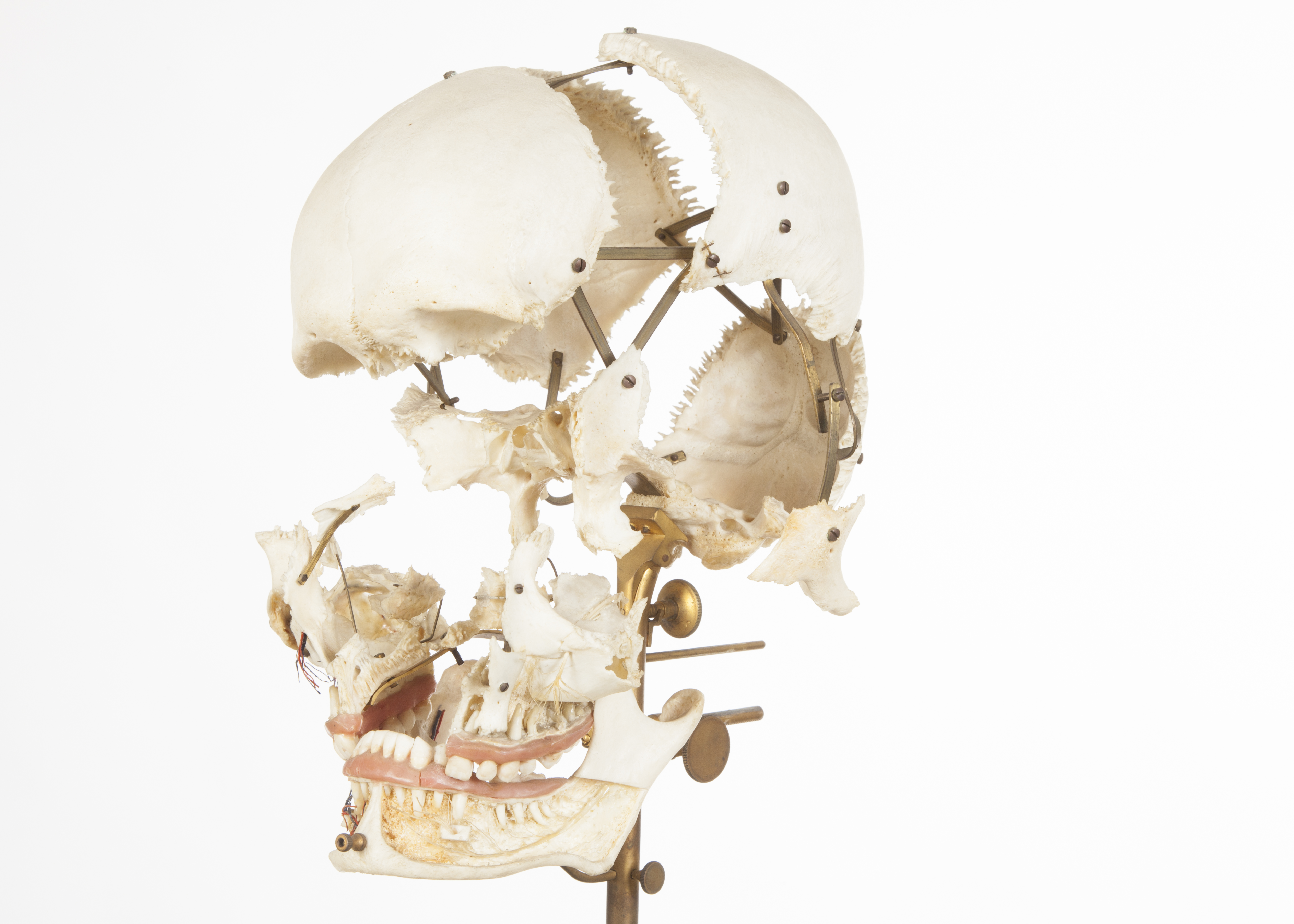 Beauchene skull Beauchene skull
Bone with wax, brass connections and wires. Mid to late 19th century. Maison Tramond
Description: Young adult skull. Skull separated on suture lines and joints, demonstrating the anatomical relations between the cranial and facial bones. The anterior cortical bone on the maxillae and mandible has been removed in order to expose the inferior and superior alveolar nerves, the mental foramen and the roots of the permanent dentition. The mental foramen, hosting the mental nerve, is an important landmark in dentistry. The infraorbital nerves are also visible, as well as the blood supply to the face on the right side. The gums are modelled in pink wax.

|
|
|
|
|
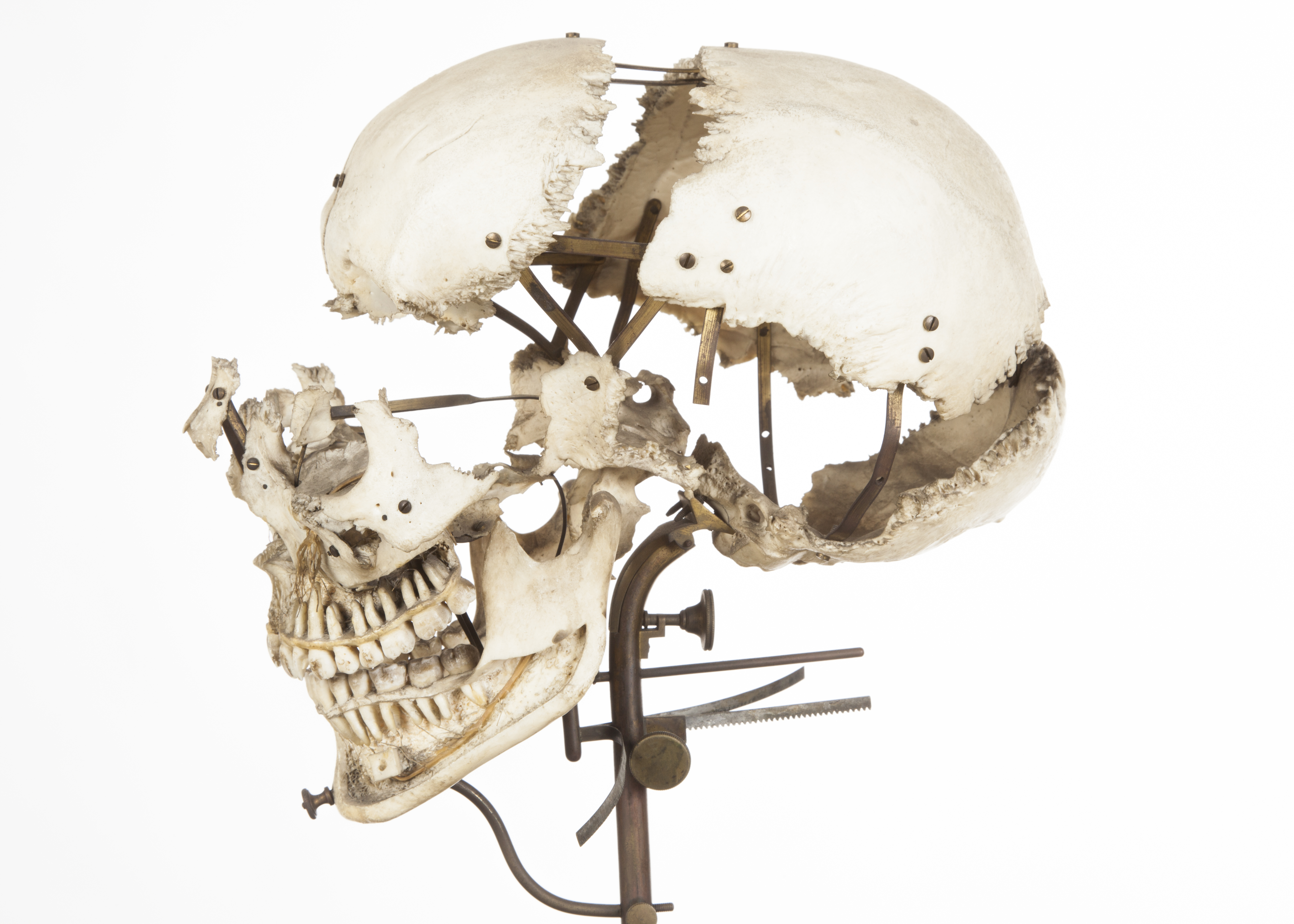 Beauchene skull Beauchene skull
Bone with wax, brass connections and wires. Mid to late 19th century. Maison Tramond.
Description: Young adult skull. Skull separated along the suture lines and joints, demonstrating the anatomical relations between the cranial and facial bones. The temporal bones are missing. The cortical bone on the anterior part of the maxillae and mandible (jaw) has been cut in order to expose the inferior and superior alveolar nerves and the roots of the permanent dentition. The infraorbital nerves are also visible. The left maxillary sinus exposed (N.B. seen through area of post-mortem damage). It is in good condition, with minor dark stains from dust, and some damage to the left maxilla.

|
|
|
|
|
|


