
Submitted by Dr R. Inchingolo on Tue, 07/08/2018 - 14:01
New research by Guy Blanchard in collaboration with the LMB Cell Biology Division gives new insight into how flat layers of cell tissue develop into 3D structures
Many complex tubular organs, including kidney, lung, the intestine and several glands, form from a flat layer of cells during animal development. Failure of proper tube formation underlies severe congenital malformations such as Spina Bifida, and the failure to maintain tube architecture for instance underlies Polycystic Kidney Diseases. How tissues transition from flat 2D structures to 3D tubes is poorly understood. New research from Katja Röper’s group in the LMB’s Cell Biology Division, in collaboration with Guy Blanchard at the Department of Physiology, Development and Neuroscience at the University of Cambridge, has recently extended knowledge of this process by exploring the cellular dynamics occurring just before and during the 2D-3D transition.
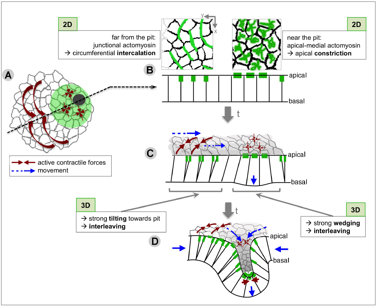
Using the fruit fly as a model organism, the formation of a simple tubular organ in the fly embryo was observed and quantitated over time. This organ starts as a roughly circular patch of cells in the surface layer of the embryo, the epidermis. It was found that these cells start to alter their shape by contracting their apical side leading to the cell becoming progressively wedge-shape. When a succession of cells in the same area change in the same way, it generates forces that result in a dimple, a future tube pit, being formed. In the rest of the tissue, cells were tilted towards the pit but gained new neighbours lying within the circumference. This helped the flow of cells towards the invagination pit, eventually leading to the formation of a tube inside the embryo.
Yara Sanchez-Corrales, in Katja’s group, carried out time-lapse experiments using confocal microscopy, to make movies that followed the changes in cell behaviours. Novel computational morphometric techniques were developed by Guy Blanchard, to extract quantitative information from the movies and identify and describe cell behaviours.
Mechanisms of organ formation are often conserved between animals, and so such analysis can inform a general understanding of tube formation. The work also provides the basis to compare cellular dynamics in a simple tubular organ to other tubular structures in which it is more difficult to obtain data at high temporal and spatial resolution. In addition, mapping cell behaviours in 2D and characterising 3D cell geometries during early stages of tube formation may help us to understand mutant situations when this process is disrupted. It could also inspire new ways of constructing artificial tissue tubes.
From the MRC Laboratory of Molecular Biology