Read more at: Celebrations for the Raffan Lab
Celebrations for the Raffan Lab
5 June 2025
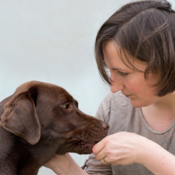
Congratulations to Dr Eleanor Raffan who is the 2025 recipient of the Blaine Award. The award is presented to veterinarians who are not client facing (e.g. pathology, clinical laboratory) for contributions to the advancement of small animal science in aspects of non-client-facing sciences, including, but not limited to...
 Malcolm Neil, Sarrah Fawcett, Cecilia Brassett & Chris Smith")