Read more at: Congratulations to Team OccuSense
Congratulations to Team OccuSense
15 August 2025
Congratulations to Enoch Alex, a PhD student member of the Raffan Lab , who was a key member of a team that won the Market Matchmakers Award at the recent Crick x Cambridge Innovation Challenge, hosted at the Francis Crick Institute. The high-profile event challenges participants to transform scientific discoveries into...


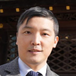
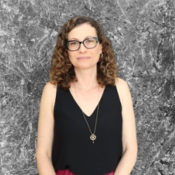
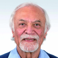
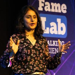
 Malcolm Neil, Sarrah Fawcett, Cecilia Brassett & Chris Smith")