skip to content

Department of Physiology, Development and Neuroscience


Welcome to the Franze Lab


Key aspects in the development of the central nervous system (CNS) include the formation of neuronal axons, their subsequent growth and guidance through thick layers of nervous tissue, and the folding of the brain. All these processes involve motion and must thus be driven by forces. However, while our understanding of the biochemical and molecular control of these processes is increasing rapidly, the contribution of mechanics remains poorly understood. Cell motion is also crucially involved in CNS pathologies such as foreign body reactions, in which activated glial cells migrate towards and encapsulate implants (e.g., electrodes), and the failing regeneration of neurons after CNS (e.g., spinal cord) injuries. Repair can currently not be promoted. So far, research has - without any major breakthrough - mainly focused on chemical signals impeding and promoting neuronal (re)growth.

We are taking an interdisciplinary approach and investigate how cellular forces, local cell and tissue compliance and cellular mechanosensitivity contribute to CNS development and disease. Methods we are exploiting include atomic force microscopy, traction force microscopy, custom-built simple and complex compliant cell culture substrates, optical microscopy including confocal laser scanning microscopy and cell biological techniques. We have shown, for example, that nervous tissue is mechanically highly heterogeneous. Furthermore, we found that neurons constantly exert forces on their environment and that both neurons and glial cells respond to mechanical cues such as tissue stiffness. Understanding how and when CNS cells actively exert forces and respond to their mechanical environment will shed new light on CNS development, and it could eventually lead to novel biomedical approaches to treat or circumvent pathologies that involve mechanical signalling.

Research

The mechanobiology of nervous system development and pathology.

Developmental biology, biophysics, fun projects.

Atomic force microscopy.


Main sources of funding: ERC, BBSRC


Main collaborators

Christoph Ballestrem (Centre for Cell Matrix Research, University of Manchester)
Kevin Chalut (Physics and SCRI, Cambridge)
James Fawcett (Brain Repair Centre, Cambridge)
Christine Holt (PDN, Cambridge)


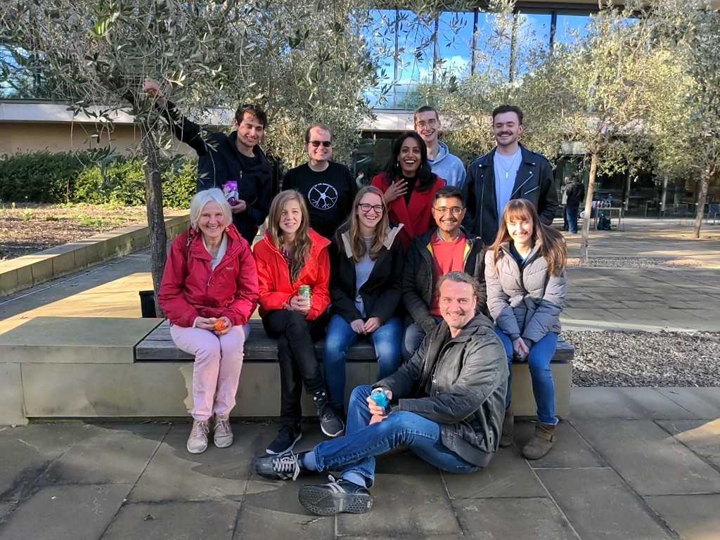
Research Team

Julia Becker (Postdoc)
Eva Kreysing (Postdoc)
Ross McGinn (Postdoc)
Rachel McKeown (PhD student)
Katrin Mooslehner (Molecular biology expert)
Sudipta Mukherjee (PhD student)
Jana Sipkova (Postdoc)
Liz Williams (lab manager)
Alex Winkel (AFM expert)


Previous Lab Members 

Andrea Dimitracopoulos (Postdoc)
Damiano Giuseppe Barone (PhD student, shared with James Fawcett)
Francesco Barone (Master's student)
Lars Bollmann (Master's student)
Alejandro Carnicer (PhD student, co-supervisor with James Fawcett)
Ivan Dimov (PhD student)
Sarah Foster (PhD student)
Hélène Gautier (postdoc)
Ryan Greenhalugh (PhD student)
Kathrin Holtzmann (PhD student, Physics/VetSchool)
Max Jacobs (PhD student)
David Koser (PhD student)
Emad Moeendarbary (postdoc)
Eva Pillai (PhD student)
Rasha Rezk (postdoc)
Sara Rolle (Masters student)
Rajesh Shahapure (postdoc)
Graham Sheridan (postdoc)
Joy Thompson (PhD student)
Elke Ulbricht (postdoc)
Omer Wagner (postdoc)
Isabell Weber (postdoc)
Vanessa Sokleva (PhD student)

Publications

Key publications: 

Jakobs MAH, Zemel A, Franze K: Unrestrained growth of correctly oriented microtubules instructs axonal microtubule orientation. Elife 11:e77608 (2022)

Rheinlaender J, Dimitracopoulos A, Wallmeyer B, Kronenberg NM, Chalut KJ, Gather MC, Betz T, Charras G, Franze K: Cortical cell stiffness is independent of substrate mechanics. Nature Materials 19:10191025 (2020)

Jakobs MAH, Dimitracopoulos A, Franze K: KymoButler, a deep learning software for automated kymograph analysis. eLife 8:e42288 (2019)

Thompson AJ, Pillai EK, Dimov IB, Foster SK, Holt CE, Franze K: Rapid changes in tissue mechanics regulate cell behaviour in the developing embryonic brain. eLife 8:e39356 (2019)

Barriga EH, Franze K, Charras G, Mayor R: Tissue stiffening coordinates morphogenesis by triggering collective cell migration in vivo. Nature doi:10.1038/nature25742 (2018)

Moeendarbary E, Weber IP, Sheridan GK, Koser DE, Solemane S, Haenzie B, Bradbury EJ, Fawcett J, Franze K: The soft mechanical signature of glial scars in the central nervous system. Nature Communications 8:14787 (2017)

Koser DE, Thompson AJ, Foster SK, Dwivedy A, Pillai EK, Sheridan GK, Svoboda H, Viana M, Costa LdF, Guck J, Holt CE, Franze K: Mechanosensing is critical for axon growth in the developing brain. Nature Neuroscience 19(12):1592-1598 (2016)

MacDonald RB, Randlett O, Oswald J, Yoshimatsu T, Franze K*, Harris WA*: Muller Glia Provide Essential Tensile Strength to the Developing Retina. Journal of Cell Biology 210(7):1075-1083 (2015)

Pagliara S*, Franze K*, McClain CR, Wylde G, Fisher CL, Franklin RJM, Kabla AJ, Keyser UF, Chalut KJ: Auxetic nuclei in embryonic stem cells exiting pluripotency. Nature Materials 13:638-644 (2014)

Hardie RC and Franze K: Photomechanical responses in Drosophila photoreceptors. Science 338(6104):260-263 (2012)

Franze K, Grosche J, Skatchkov SN, Schinkinger S, Foja C, Schild D, Uckermann O, Travis K, Reichenbach A, Guck J: Müller cells are living optical fibers in the vertebrate retina. PNAS 104(20):8287-8292 (2007)

Teaching and Supervisions

Professor of Neuronal Mechanics
Fellow of St. John's College
Kristian  Franze

Contact Details

Department of Physiology, Development and Neuroscience
University of Cambridge
Downing Street
CambridgeCB2 3DY, UK
Email: kf284@cam.ac.uk
Phone: +44 (0)1223 3-33761
Email address: